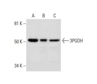
3PGDH Antikörper (B-1): sc-390610
- 3PGDH Antikörper B-1 ist ein Maus monoklonales IgG1 κ 3PGDH Antikörper, verwendet in 1 wissenschaftlichen Veröffentlichungen, in einer Menge von 200 µg/ml
- gezogen gegen die Aminosäuresequenz 260-533 lokalisiert am C-terminus von 3PGDH aus der Spezies human
- 3PGDH Antikörper (B-1) ist empfohlen für die Detektion von 3PGDH aus der Spezies mouse, rat und human per WB, IP, IF und ELISA
- Anti-3PGDH Antikörper (B-1) ist erhältlich als Konjugat mit Agarose für IP; HRP für WB, IHC(P) und ELISA; und entweder mit Phycoerythrin oder FITC für IF, IHC(P) und FCM
- auch erhältlich als Konjugat mit Alexa Fluor® 488, Alexa Fluor® 546, Alexa Fluor® 594 oder Alexa Fluor® 647 für IF, IHC(P) und FCM
- auch erhältlich als Konjugat mit Alexa Fluor® 680 oder Alexa Fluor® 790 für WB (NIR), IF und FCM
- m-IgG Fc BP-HRP, 1 BP-HRP">m-IgG1 BP-HRP und m-IgGκ BP-HRP sind die bevorzugten sekundären Nachweisreagenzien für 3PGDH Antikörper (B-1) für WB-Anwendungen. Diese Reagenzien werden jetzt in Bündeln mit 3PGDH Antikörper (B-1) angeboten(siehe Bestellinformationen unten).
Der 3PGDH-Antikörper (B-1) ist ein monoklonaler Maus IgG1 κ 3PGDH-Antikörper (auch als SERA-Antikörper, Phosphoglycerat-Dehydrogenase-Antikörper, D-3-Phosphoglycerat-Dehydrogenase-Antikörper, 2-Oxoglutarat-Reduktase-Antikörper, Malat-Dehydrogenase-Antikörper, PGDH-Antikörper, SERA-Antikörper, PDG-Antikörper, 3-Phosphoglycerat-Dehydrogenase-Antikörper, PHGDHD-Antikörper, PGDH3-Antikörper, NLS1-Antikörper, PGAD-Antikörper, NLS-Antikörper oder PGD-Antikörper bezeichnet), der das 3PGDH-Protein von Maus-, Ratte- und menschlicher Herkunft mittels WB, IP, IF und ELISA detektiert. Der 3PGDH-Antikörper (B-1) ist sowohl in nicht konjugierter Form als auch in mehreren konjugierten Formen des Anti-3PGDH-Antikörpers erhältlich, darunter Agarose, HRP, PE, FITC und mehrere Alexa Fluor®-Konjugate. Die Überlebens- und Entwicklung zentraler Neuronen erfordern die Versorgung mit trophischen Faktoren durch Gliazellen. Die trophischen Wirkungen von Gliazellen auf Purkinje-Neuronen werden durch L-Serin und Glycin vermittelt, die glia-abgeleitete trophische Faktoren sind, die durch 3PGDH synthetisiert werden. Das 3PGDH-Protein ist 544 Aminosäuren lang. Zwei unterschiedliche mRNA-Transkripte, die für das 3PGDH-Protein in normalen menschlichen Geweben codieren, sind das dominante 2,1 kb mRNA, das hauptsächlich in Prostata, Hoden, Eierstock, Gehirn, Leber, Niere und Bauchspeicheldrüse hoch und schwach in Thymus, Colon und Herz exprimiert wird, und das 710 bp mRNA, das hauptsächlich in Herz und Skelettmuskel hoch exprimiert wird. 3PGDH wird je nach Gewebespezifität und zellulärem Proliferationsstatus auf transcriptioneller Ebene reguliert. Das 3PGDH-Protein ist auch in adulten und fetalen Gehirngeweben hoch exprimiert. 3PGDH-Protein spielt eine wichtige Rolle bei Stoffwechsel, Entwicklung und Funktion des zentralen Nervensystems und sein Mangel ist ein behandelbarer angeborener Fehler, der die L-Serin-Biosynthese beeinträchtigt und der durch angeborene Mikrozephalie, psychomotorische Retardierung und Krampfanfälle charakterisiert ist.
Alexa Fluor® ist ein Markenzeichen von Molecular Probes Inc., OR., USA
LI-COR® und Odyssey® sind Markenzeichen von LI-COR Biosciences
3PGDH Antikörper (B-1) Literaturhinweise:
- 3-Phosphoglycerat-Dehydrogenase, ein Schlüsselenzym für die l-Serin-Biosynthese, wird bevorzugt in der radialen Glia/Astrozyten-Linie und den olfaktorischen Glia-Scheiden im Mäusegehirn exprimiert. | Yamasaki, M., et al. 2001. J Neurosci. 21: 7691-704. PMID: 11567059
- Die Aktivierung der 3-Phosphoglycerat-Dehydrogenase durch L-Methionin. | Slaughter, JC. 1970. FEBS Lett. 7: 245-247. PMID: 11947482
- Die Expression von 3-Phosphoglycerat-Dehydrogenase wird durch HOXA10 in murinem Endometrium und menschlichen Endometriumzellen reguliert. | Du, H., et al. 2010. Reproduction. 139: 237-45. PMID: 19778996
- D-Serin in Glia und Neuronen wird aus 3-Phosphoglycerat-Dehydrogenase gewonnen. | Ehmsen, JT., et al. 2013. J Neurosci. 33: 12464-9. PMID: 23884950
- Identifizierung eines niedermolekularen Inhibitors der 3-Phosphoglycerat-Dehydrogenase zur gezielten Bekämpfung der Serin-Biosynthese bei Krebserkrankungen. | Mullarky, E., et al. 2016. Proc Natl Acad Sci U S A. 113: 1778-83. PMID: 26831078
- Erhöhte Anfälligkeit für oxidativen Stress und Induktion einer entzündlichen Genexpression in Fibroblasten mit 3-Phosphoglycerat-Dehydrogenase-Mangel. | Hamano, M., et al. 2018. FEBS Open Bio. 8: 914-922. PMID: 29928571
- Azacoccon E hemmt das Wachstum von Krebszellen, indem es auf die 3-Phosphoglycerat-Dehydrogenase wirkt. | Guo, J., et al. 2019. Bioorg Chem. 87: 16-22. PMID: 30852233
- Die Hemmung der 3-Phosphoglycerat-Dehydrogenase (PHGDH) durch Indolamide hemmt die De-novo-Serinsynthese in Krebszellen. | Mullarky, E., et al. 2019. Bioorg Med Chem Lett. 29: 2503-2510. PMID: 31327531
- Determinanten der Substratspezifität der D-3-Phosphoglycerat-Dehydrogenase. Umwandlung des M. tuberculosis-Enzyms von einem Enzym, das α-Ketoglutarat nicht als Substrat verwendet, zu einem, das dies tut. | Xu, XL. and Grant, GA. 2019. Arch Biochem Biophys. 671: 218-224. PMID: 31344342
- Metabolische Kompensation aktiviert überlebensfördernde mTORC1-Signale bei Hemmung der 3-Phosphoglycerat-Dehydrogenase in Osteosarkomen. | Rathore, R., et al. 2021. Cell Rep. 34: 108678. PMID: 33503424
Bestellinformation
| Produkt | Katalog # | EINHEIT | Preis | ANZAHL | Favoriten | |
3PGDH Antikörper (B-1) | sc-390610 | 200 µg/ml | CNY2422.00 | |||
3PGDH (B-1): m-IgG Fc BP-HRP Bundle | sc-530202 | 200 µg Ab; 10 µg BP | CNY2715.00 | |||
3PGDH (B-1): m-IgGκ BP-HRP Bundle | sc-523603 | 200 µg Ab, 40 µg BP | CNY2715.00 | |||
3PGDH (B-1): m-IgG1 BP-HRP Bundle | sc-543926 | 200 µg Ab; 20 µg BP | CNY2715.00 | |||
3PGDH Antikörper (B-1) AC | sc-390610 AC | 500 µg/ml, 25% agarose | CNY3189.00 | |||
3PGDH Antikörper (B-1) HRP | sc-390610 HRP | 200 µg/ml | CNY2422.00 | |||
3PGDH Antikörper (B-1) FITC | sc-390610 FITC | 200 µg/ml | CNY2527.00 | |||
3PGDH Antikörper (B-1) PE | sc-390610 PE | 200 µg/ml | CNY2625.00 | |||
3PGDH Antikörper (B-1) Alexa Fluor® 488 | sc-390610 AF488 | 200 µg/ml | CNY2738.00 | |||
3PGDH Antikörper (B-1) Alexa Fluor® 546 | sc-390610 AF546 | 200 µg/ml | CNY2738.00 | |||
3PGDH Antikörper (B-1) Alexa Fluor® 594 | sc-390610 AF594 | 200 µg/ml | CNY2738.00 | |||
3PGDH Antikörper (B-1) Alexa Fluor® 647 | sc-390610 AF647 | 200 µg/ml | CNY2738.00 | |||
3PGDH Antikörper (B-1) Alexa Fluor® 680 | sc-390610 AF680 | 200 µg/ml | CNY2738.00 | |||
3PGDH Antikörper (B-1) Alexa Fluor® 790 | sc-390610 AF790 | 200 µg/ml | CNY2738.00 |

