")
VCAM-1 Antikörper (E-10): sc-13160
- VCAM-1 Antikörper E-10 ist ein Maus monoklonales IgG1 κ VCAM-1 Antikörper, verwendet in 201 wissenschaftlichen Veröffentlichungen, in einer Menge von 200 µg/ml
- gezogen gegen die Aminosäuresequenz 25-300 von VCAM-1 aus der Spezies human
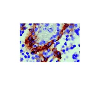
- VCAM-1 Antikörper (E-10) ist empfohlen für die Detektion von VCAM-1 aus der Spezies mouse, rat und human per WB, IP, IF, IHC(P), FCM und ELISA
- Anti-VCAM-1 Antikörper (E-10) ist erhältlich als Konjugat mit Agarose für IP; HRP für WB, IHC(P) und ELISA; und entweder mit Phycoerythrin oder FITC für IF, IHC(P) und FCM
- auch erhältlich als Konjugat mit Alexa Fluor® 488, Alexa Fluor® 546, Alexa Fluor® 594 oder Alexa Fluor® 647 für IF, IHC(P) und FCM
- auch erhältlich als Konjugat mit Alexa Fluor® 680 oder Alexa Fluor® 790 für WB (NIR), IF und FCM
- auch konjugiert erhältlich mit Alexa Fluor® 405 für IF, IHC(P) und FCM
- m-IgG Fc BP-HRP, 1 BP-HRP">m-IgG1 BP-HRP und m-IgGκ BP-HRP sind die bevorzugten sekundären Nachweisreagenzien für VCAM-1 Antikörper (E-10) für WB- und IHC(P)-Anwendungen. Diese Reagenzien werden jetzt in Bündeln mit VCAM-1 Antikörper (E-10) angeboten(siehe Bestellinformationen unten).

Direktverknüpfungen
Der VCAM-1-Antikörper (E-10) ist ein monoklonaler IgG1-Kappa-Leichtketten-Antikörper der Maus, der gegen die Aminosäuren 25-300 des humanen vaskulären Zelladhäsionsmoleküls 1 (VCAM-1) gerichtet ist. VCAM-1, auch als CD106 bekannt, ist ein Transmembran-Glykoprotein, das zur Immunglobulin-Superfamilie gehört und hauptsächlich auf aktivierten Endothelzellen exprimiert wird. Strukturell besteht VCAM-1 aus sieben Immunglobulin-ähnlichen Domänen, die für die Bindung an Integrine wie VLA-4 (α4β1) auf Leukozyten unerlässlich sind und deren Adhäsion und Transmigration durch das Endothel während Immunreaktionen erleichtern. Posttranslationale Modifikationen wie die N-verknüpfte Glykosylierung sind für die korrekte Faltung, Stabilität und Funktion von VCAM-1 von entscheidender Bedeutung und beeinflussen die Interaktion mit Integrinen und die gesamten Zelladhäsionsprozesse. Die Expression von VCAM-1 wird durch entzündungsfördernde Zytokine wie Interleukin-1 (IL-1), Tumornekrosefaktor-alpha (TNF-α) und Lipopolysaccharid (LPS) hochreguliert, wodurch VCAM-1 mit verschiedenen entzündlichen Erkrankungen wie Atherosklerose und rheumatoider Arthritis in Verbindung gebracht wird. Der Anti-VCAM-1-Antikörper (E-10) reagiert mit VCAM-1 von Mäusen, Ratten und Menschen und eignet sich für Anwendungen wie Western Blot (WB), Immunpräzipitation (IP), Immunfluoreszenz (IF), Immunhistochemie mit in Paraffin eingebetteten Schnitten (IHCP), Durchflusszytometrie (FCM) und Enzyme-linked Immunosorbent Assay (ELISA). Der VCAM-1 (E-10)-Antikörper dient als wertvolles Instrument zur Untersuchung der Endothelaktivierung, der Leukozytenadhäsion und der Mechanismen, die entzündlichen Prozessen zugrunde liegen. Das Verständnis der VCAM-1-Struktur und der posttranslationalen Modifikationen ist wichtig für die Entwicklung therapeutischer Strategien gegen Krankheiten, die mit einem abnormalen Immunzelltransport und einer vaskulären Dysfunktion einhergehen.
Alexa Fluor® ist ein Markenzeichen von Molecular Probes Inc., OR., USA
LI-COR® und Odyssey® sind Markenzeichen von LI-COR Biosciences
VCAM-1 Antikörper (E-10) Literaturhinweise:
- Tumor-Nekrose-Faktor in Kombination mit IL-4 oder IFN-gamma erhöht selektiv die Adhäsionsfähigkeit von Endothelzellen für T-Zellen. Der Beitrag von Vascular Cell Adhesion Molecule-1-abhängigen und -unabhängigen Bindungsmechanismen. | Thornhill, MH., et al. 1991. J Immunol. 146: 592-8. PMID: 1702807
- Vaskuläres Zelladhäsionsmolekül-1 (VCAM-1) – ein wachsendes Verständnis seiner Rolle bei der Tumorentstehung und Metastasierung. | Schlesinger, M. and Bendas, G. 2015. Int J Cancer. 136: 2504-14. PMID: 24771582
- Neue Rollen des vaskulären Zelladhäsionsmoleküls-1 (VCAM-1) bei immunologischen Störungen und Krebs. | Kong, DH., et al. 2018. Int J Mol Sci. 19: PMID: 29614819
- Das lipidinduzierte endotheliale vaskuläre Zelladhäsionsmolekül 1 fördert die Pathogenese der nichtalkoholischen Steatohepatitis. | Furuta, K., et al. 2021. J Clin Invest. 131: PMID: 33476308
- Neuronales Zelladhäsionsmolekül (NCAM) als quantitativer Marker beim synaptischen Umbau. | Jørgensen, OS. 1995. Neurochem Res. 20: 533-47. PMID: 7643959
- Die Nutzung des Vase-Mini-Exons durch NCAM ist nicht auf Tumore neuroektodermalen Ursprungs beschränkt. | Patel, K., et al. 1993. Int J Cancer. 54: 772-7. PMID: 8325706
- Das Modell der Kallmann-Syndrom-Variante (KSV) der Schizophrenien. | Cowen, MA. and Green, M. 1993. Schizophr Res. 9: 1-10. PMID: 8461265
- Adhäsionsmoleküle von Endothelzellen und Leukozyten. | Bevilacqua, MP. 1993. Annu Rev Immunol. 11: 767-804. PMID: 8476577
- Mikrometastasen im Knochenmark bei einem Patienten mit lokalisiertem Wilms-Tumor. | Dominici, C., et al. 1996. Med Pediatr Oncol. 26: 125-8. PMID: 8531850
- Strukturelle Voraussetzungen für die Bindung des mukosalen vaskulären Adressins an seinen Lymphozytenrezeptor Alpha 4 Beta 7. Gemeinsame Themen bei den Interaktionen zwischen Integrin und Ig-Familie. | Briskin, MJ., et al. 1996. J Immunol. 156: 719-26. PMID: 8543825
- Antikörper gegen Proteinase 3 vermitteln die Expression des vaskulären Zelladhäsionsmoleküls 1 (VCAM-1). | Mayet, WJ., et al. 1996. Clin Exp Immunol. 103: 259-67. PMID: 8565309
- Entwicklungsbedingte Kontrolle der N-CAM-Expression durch Hox- und Pax-Genprodukte. | Edelman, GM. and Jones, FS. 1995. Philos Trans R Soc Lond B Biol Sci. 349: 305-12. PMID: 8577842
Bestellinformation
| Produkt | Katalog # | EINHEIT | Preis | ANZAHL | Favoriten | |
VCAM-1 Antikörper (E-10) | sc-13160 | 200 µg/ml | CNY2422.00 | |||
VCAM-1 (E-10): m-IgG Fc BP-HRP Bundle | sc-528279 | 200 µg Ab; 10 µg BP | CNY2715.00 | |||
VCAM-1 (E-10): m-IgGκ BP-HRP Bundle | sc-520618 | 200 µg Ab, 40 µg BP | CNY2715.00 | |||
VCAM-1 (E-10): m-IgG1 BP-HRP Bundle | sc-542849 | 200 µg Ab; 20 µg BP | CNY2715.00 | |||
VCAM-1 Antikörper (E-10) AC | sc-13160 AC | 500 µg/ml, 25% agarose | CNY3189.00 | |||
VCAM-1 Antikörper (E-10) HRP | sc-13160 HRP | 200 µg/ml | CNY2422.00 | |||
VCAM-1 Antikörper (E-10) FITC | sc-13160 FITC | 200 µg/ml | CNY2527.00 | |||
VCAM-1 Antikörper (E-10) PE | sc-13160 PE | 200 µg/ml | CNY2625.00 | |||
VCAM-1 Antikörper (E-10) Alexa Fluor® 488 | sc-13160 AF488 | 200 µg/ml | CNY2738.00 | |||
VCAM-1 Antikörper (E-10) Alexa Fluor® 546 | sc-13160 AF546 | 200 µg/ml | CNY2738.00 | |||
VCAM-1 Antikörper (E-10) Alexa Fluor® 594 | sc-13160 AF594 | 200 µg/ml | CNY2738.00 | |||
VCAM-1 Antikörper (E-10) Alexa Fluor® 647 | sc-13160 AF647 | 200 µg/ml | CNY2738.00 | |||
VCAM-1 Antikörper (E-10) Alexa Fluor® 680 | sc-13160 AF680 | 200 µg/ml | CNY2738.00 | |||
VCAM-1 Antikörper (E-10) Alexa Fluor® 790 | sc-13160 AF790 | 200 µg/ml | CNY2738.00 | |||
VCAM-1 Antikörper (E-10) Alexa Fluor® 405 | sc-13160 AF405 | 200 µg/ml | CNY2738.00 |