")
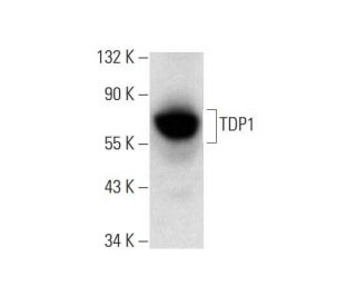
: sc-365674. Western Blot Analyse der TDP1 Expression in Ramos-Ganzzell-Lysat.")
: sc-365674. Immunoperoxidasefärbung von in Formalin fixiertem, in Paraffin eingebettetem menschlichem Schilddrüsengewebe mit zytoplasmatischer Färbung der Drüsenzellen.")
: sc-365674. Nahinfrarot-Westernblot-Analyse der TDP1 Expression in Ramos-Vollzell-Lysat. Blockiert mit UltraCruz® Blocking Reagent: sc-516214. Verwendetes Detektionsreagenz: m-IgGκ BP-CFL 647: sc-516179.")
TDP1 Antikörper (C-3): sc-365674
- TDP1 Antikörper C-3 ist ein Maus monoklonales IgG1 κ TDP1 Antikörper, verwendet in 10 wissenschaftlichen Veröffentlichungen, in einer Menge von 200 µg/ml
- gezogen gegen die Aminosäuresequenz 309-608 lokalisiert am C-terminus von TDP1 aus der Spezies human
- TDP1 Antikörper (C-3) ist empfohlen für die Detektion von TDP1 aus der Spezies human per WB, IP, IF, IHC(P) und ELISA
- Anti-TDP1 Antikörper (C-3) ist erhältlich als Konjugat mit Agarose für IP; HRP für WB, IHC(P) und ELISA; und entweder mit Phycoerythrin oder FITC für IF, IHC(P) und FCM
- auch erhältlich als Konjugat mit Alexa Fluor® 488, Alexa Fluor® 546, Alexa Fluor® 594 oder Alexa Fluor® 647 für IF, IHC(P) und FCM
- auch erhältlich als Konjugat mit Alexa Fluor® 680 oder Alexa Fluor® 790 für WB (NIR), IF und FCM
- m-IgG Fc BP-HRP und m-IgG1 BP-HRP sind die bevorzugten sekundären Nachweisreagenzien für TDP1 Antikörper (C-3) for WB and IHC(P) applications. Diese Reagenzien werden jetzt in Bündeln mit TDP1 Antikörper (C-3) angeboten(siehe Bestellinformationen unten).

Direktverknüpfungen
Siehe auch...
Die Tyrosyl-DNA-Phosphodiesterase 1 (TDP1), ein DNA-Reparatur-Enzym, katalysiert die Hydrolyse von Phophodiester-Bindungen zwischen Tyrosin-Resten und DNA-3'-Phosphaten. Darüber hinaus entfernt TDP1 Glycolat aus einzelsträngiger DNA mit einem 3'-Phosphoglycolat, was auf eine Rolle bei der Reparatur von durch freie Radikale vermittelten DNA-Doppelstrangbrüchen hinweist. Ein einzigartiges HKD-Signaturmotiv mit hochkonservierten Lysin- und Histidin-Resten, das in TDP1 vorhanden ist, stellt das Enzym in eine eigene Klasse innerhalb der Phospholipase-D-Superfamilie. Die hydrolytische Reaktion, die von TDP1 katalysiert wird, erfolgt durch eine Phosphoryl-Transfer-Reaktion, die allen Mitgliedern der PLD-Superfamilie gemeinsam ist. Verlustfunktionsmutationen in TDP1 können zu einer spinozerebellären Ataxie mit axonaler Neuropathie führen, indem sie die DNA-Transkription stören oder Apoptose in postmitotischen Neuronen induzieren. Der TDP1-Antikörper (C-3) ist ein IgG1 κ-muriner Monoklonaler TDP1-Antikörper (auch als TDP1-Antikörper bezeichnet), der das TDP1-Protein menschlichen Ursprungs mittels WB, IP, IF, IHC (P) und ELISA detektiert. Der TDP1-Antikörper (C-3) ist sowohl in Form des nicht konjugierten Anti-TDP1-Antikörpers als auch in mehreren konjugierten Formen des Anti-TDP1-Antikörpers, einschließlich Agarose, HRP, PE, FITC und mehreren Alexa Fluor®-Konjugaten, erhältlich.
Alexa Fluor® ist ein Markenzeichen von Molecular Probes Inc., OR., USA
LI-COR® und Odyssey® sind Markenzeichen von LI-COR Biosciences
TDP1 Antikörper (C-3) Literaturhinweise:
- Die Tyrosyl-DNA-Phosphodiesterase Tdp1 ist ein Mitglied der Phospholipase-D-Superfamilie. | Interthal, H., et al. 2001. Proc Natl Acad Sci U S A. 98: 12009-14. PMID: 11572945
- Umwandlung von Phosphoglykolat in Phosphat-Termini an 3'-Überhängen von DNA-Doppelstrangbrüchen durch die menschliche Tyrosyl-DNA-Phosphodiesterase hTdp1. | Inamdar, KV., et al. 2002. J Biol Chem. 277: 27162-8. PMID: 12023295
- Mutation von TDP1, das für ein Topoisomerase I-abhängiges Enzym zur Reparatur von DNA-Schäden kodiert, bei spinozerebellarer Ataxie mit axonaler Neuropathie. | Takashima, H., et al. 2002. Nat Genet. 32: 267-72. PMID: 12244316
- Einblicke in die Substratbindung und den katalytischen Mechanismus der menschlichen Tyrosyl-DNA-Phosphodiesterase (Tdp1) aus Vanadat- und Wolframat-inhibierten Strukturen. | Davies, DR., et al. 2002. J Mol Biol. 324: 917-32. PMID: 12470949
Bestellinformation
| Produkt | Katalog # | EINHEIT | Preis | ANZAHL | Favoriten | |
TDP1 Antikörper (C-3) | sc-365674 | 200 µg/ml | CNY2422.00 | |||
TDP1 (C-3): m-IgG Fc BP-HRP Bundle | sc-527389 | 200 µg Ab; 10 µg BP | CNY2715.00 | |||
TDP1 (C-3): m-IgG1 BP-HRP Bundle | sc-532762 | 200 µg Ab; 20 µg BP | CNY2715.00 | |||
TDP1 Antikörper (C-3) AC | sc-365674 AC | 500 µg/ml, 25% agarose | CNY3189.00 | |||
TDP1 Antikörper (C-3) HRP | sc-365674 HRP | 200 µg/ml | CNY2422.00 | |||
TDP1 Antikörper (C-3) FITC | sc-365674 FITC | 200 µg/ml | CNY2527.00 | |||
TDP1 Antikörper (C-3) PE | sc-365674 PE | 200 µg/ml | CNY2625.00 | |||
TDP1 Antikörper (C-3) Alexa Fluor® 488 | sc-365674 AF488 | 200 µg/ml | CNY2738.00 | |||
TDP1 Antikörper (C-3) Alexa Fluor® 546 | sc-365674 AF546 | 200 µg/ml | CNY2738.00 | |||
TDP1 Antikörper (C-3) Alexa Fluor® 594 | sc-365674 AF594 | 200 µg/ml | CNY2738.00 | |||
TDP1 Antikörper (C-3) Alexa Fluor® 647 | sc-365674 AF647 | 200 µg/ml | CNY2738.00 | |||
TDP1 Antikörper (C-3) Alexa Fluor® 680 | sc-365674 AF680 | 200 µg/ml | CNY2738.00 | |||
TDP1 Antikörper (C-3) Alexa Fluor® 790 | sc-365674 AF790 | 200 µg/ml | CNY2738.00 |