")
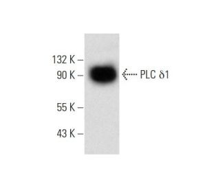
: sc-365812. Western Blot Analyse der PLC δ1 Expression in F9-Vollzell-Lysat.")
: sc-365812. Immunoperoxidasefärbung von in Formalin fixiertem, in Paraffin eingebettetem menschlichem Zwölffingerdarmgewebe mit zytoplasmatischer Färbung von Drüsenzellen.")
: sc-365812. Western Blot Analyse der PLC δ1 Expression in NIH/3T3 (A), BC3H1 (B), KNRK (C) und A-10 (D) Vollzell-Lysaten.")
PLC δ1 Antikörper (A-4): sc-365812
- PLC δ1 Antikörper (A-4) ist ein Maus monoklonales IgG1 κ, verwendet in 1 wissenschaftlichen Veröffentlichungen, in einer Menge von 200 µg/ml
- gezogen gegen die Aminosäuresequenz 1-140 lokalisiert am N-terminus von PLC δ1 aus der Spezies human
- Empfohlen für die Detektion von PLC δ1 aus der Spezies mouse, rat und human per WB, IP, IF, IHC(P) und ELISA
- m-IgG Fc BP-HRP, m-IgG1 BP-HRP und m-IgGκ BP-HRP sind die empfohlenen Detektionsreagenzien für PLC δ1 Antikörper (A-4) für WB and IHC(P) Experimente. Diese Reagenzien werden nun auch als Bundle angeboten, zusammen mit PLC δ1 Antikörper (A-4) (Bestellinformation unten stehend).

Phosphoinositid-spezifische Phospholipase C (PLC) spielt eine entscheidende Rolle bei der Initiierung der receptorvermittelten Signaltransduktion durch die Generierung der beiden Sekundärmessenger Inositol 1,4,5-Triphosphat (IP3) und Diacylglycerol (DAG) aus Phosphatidylinositol 4,5-Bisphosphat. Es gibt mehrere Säugetier-PLC-Proteine, einschließlich PLC β1, PLC β2, PLC β3, PLC β4, PLCγ1, PLCγ2, PLC δ1, PLC δ3, PLC δ4 und PLCe. PLC δ1, ein Calciumsignalverstärker, wird durch ein atypisches GTP-bindendes Protein aktiviert und fungiert als Effektor für GTP-bindendes Protein Transglutaminase II-vermittelte Oxytocin-Rezeptor- und α1B-Adrenorezeptor-Signalisierung. PLC δ1 ist hoch in Gehirn, Herz, Lunge und Hoden exprimiert und wird abnorm in autopsierten Gehirnen mit Alzheimer-Krankheit (AD) angereichert, was darauf hindeutet, dass es eine Rolle in der Pathologie von AD spielen könnte. Sowohl PLC δ3 als auch PLC δ4 enthalten mehrere funktionelle Domänen, durch die sie Calcium als Co-Faktor binden und die Bildung von DAG und IP3 katalysieren, was eine essentielle Rolle in der Signaltransduktion spielt. PLC δ4 ist hoch in Skelettmuskel- und Nierentissue sowie in kornealen Epithelzellen exprimiert, was auf eine Rolle in der Regulation von Nieren- und Augenfunktion hinweist.
Alexa Fluor® ist ein Markenzeichen von Molecular Probes Inc., OR., USA
LI-COR® und Odyssey® sind Markenzeichen von LI-COR Biosciences
PLC δ1 Antikörper (A-4) Literaturhinweise:
- Amyloid-Beta-Peptid (Abeta42) aktiviert den PLC-delta1-Promotor über die NF-kappaB-Bindestelle. | Kim, JY., et al. 2003. Biochem Biophys Res Commun. 310: 904-9. PMID: 14550290
- Mutationen in PLCδ1, die mit hereditärer Leukonychie assoziiert sind, weisen eine abweichende PIP2-hydrolytische Funktion auf. | Nomikos, M., et al. 2016. FEBS J. 283: 4502-4514. PMID: 27783455
- Die Phosphoinositidhydrolase Phospholipase C delta1 hemmt den epithelial-mesenchymalen Übergang und wird bei Darmkrebs zum Schweigen gebracht. | Xiang, Q., et al. 2019. J Cell Physiol. 234: 13906-13916. PMID: 30618183
- IQGAP1 aktiviert PLC-δ1 durch direkte Bindung und Bewegung entlang der Mikrotubuli mit DLC-1 zur Zelloberfläche. | Tanaka, M., et al. 2019. FASEB Bioadv. 1: 465-480. PMID: 32123844
- Die Gαh/Phospholipase C-δ1-Interaktion fördert den Autophagosomen-Abbau durch Aktivierung des Akt/mTORC1-Signalwegs bei metastasierendem dreifach negativem Brustkrebs. | Lin, HY., et al. 2020. Aging (Albany NY). 12: 13023-13037. PMID: 32615541
- Negative Selbstregulierung des kanonischen Transient-Rezeptor-Potentials 4 durch die spezifische Interaktion mit Phospholipase C-δ1. | Ko, J., et al. 2023. Korean J Physiol Pharmacol. 27: 187-196. PMID: 36815258
- Osmotisch evozierte PLCδ1-abhängige Translokation von ΔN-TRPV1-Kanälen in supraoptischen Neuronen der Ratte. | Haan, KD., et al. 2023. iScience. 26: 106258. PMID: 36926650
- Eine ternäre Metallbindungsstelle in der C2-Domäne der phosphoinositidspezifischen Phospholipase C-delta1. | Essen, LO., et al. 1997. Biochemistry. 36: 2753-62. PMID: 9062102
- Phosphoinositid-spezifische Phospholipase C delta1-Aktivität gegenüber mizellaren Substraten, Inositol-1,2-cyclischem Phosphat und anderen wasserlöslichen Substraten: ein sequentieller Mechanismus und allosterische Aktivierung. | Wu, Y., et al. 1997. Biochemistry. 36: 11223-33. PMID: 9287165
- Katalytische Domäne der Phosphoinositid-spezifischen Phospholipase C (PLC). Mutationsanalyse von Resten innerhalb des aktiven Zentrums und des hydrophoben Rückens von plcdelta1. | Ellis, MV., et al. 1998. J Biol Chem. 273: 11650-9. PMID: 9565585
Bestellinformation
| Produkt | Katalog # | EINHEIT | Preis | ANZAHL | Favoriten | |
PLC δ1 Antikörper (A-4) | sc-365812 | 200 µg/ml | CNY2422.00 | |||
PLC δ1 (A-4): m-IgG Fc BP-HRP Bundle | sc-537841 | 200 µg Ab; 10 µg BP | CNY2715.00 | |||
PLC δ1 (A-4): m-IgGκ BP-HRP Bundle | sc-535263 | 200 µg Ab; 40 µg BP | CNY2715.00 | |||
PLC δ1 (A-4): m-IgG1 BP-HRP Bundle | sc-545395 | 200 µg Ab; 20 µg BP | CNY2715.00 |