")
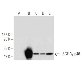
ISGF-3γ p48 항체 (H-10): sc-365893
- ISGF-3 gamma p48 항체 H-10 는 마우스 monoclonal IgG1 κ ISGF-3γ p48 항체, 13간행물에 인용, 이며 200 µg/ml으로 제공합니다.
- human origin의 ISGF-3γ p48에서 내부에 위치한 113-255 아미노산을 항원으로 사용하였습니다.
- ISGF-3 gamma p48 항체 (H-10)는 WB, IP, IF, IHC(P) and ELISA으로 human유래의 ISGF-3γ p48 를 감지하는 데에 추천한다 .
- 항-ISGF-3 gamma p48 항체(H-10)는 IP용 agarose와 결합되어 이용 가능하며, WB, IHC(P), ELISA용 HRP와 결합되어 이용 가능하며, IF, IHC(P), FCM용 phycoerythrin 또는 FITC와 결합되어 이용 가능합니다.
- WB (RGB), IF, IHC(P) 와FCM, RGB fluorescent imaging systems, such as iBright™ FL1000, FluorChem™, Typhoon, Azure and other comparable systems에 사용가능한 Alexa Fluor® 488, Alexa Fluor® 546, Alexa Fluor® 594 or Alexa Fluor® 647결합제품도 있습니다.
- WB (NIR), IF와FCM,Near-Infrared (NIR) detection systems, such as LI-COR®Odyssey®, iBright™ FL1000, FluorChem™, Typhoon, Azure and other comparable systems에 사용가능한 Alexa Fluor® 680 or Alexa Fluor® 790 결합제품도 있습니다.
- ChIP 에 사용하는 TransCruz시약(sc-365893 X, 200 µg/0.1 ml)이 있습니다.
- m-IgG Fc BP-HRP, 1 BP-HRP">m-IgG1 BP-HRP 및 m-IgGκ BP-HRP는 ISGF-3γ p48 항체(H-10) WB 및 IHC(P) 애플리케이션용입니다. 이 시약은 이제 ISGF-3γ p48 항체(H-10)와 함께 번들로 제공됩니다(아래 주문 정보 참조).

빠른 링크
더보기
ISGF-3γ p48 항체(H-10)는 IgG1 κ 마우스 단일 클론 ISGF-3 감마 p48 항체(인터페론 조절 인자 9 항체, 인터페론 자극 전사 인자 3 감마 48kDa 항체, IFN-알파 반응성 전사 인자 서브 유닛 항체로도 지정됨)입니다, 전사조절인자 ISGF3 서브유닛 감마 항체, 인터페론 자극 유전자 인자 3 감마 항체, ISGF3 P48 서브유닛 항체, ISGF-3 감마 항체, ISGF3G 항체, IRF-9 항체, ISGF3 항체 또는 P48 항체)로 인간 유래의 ISGF-3 감마 p48 단백을 검출하는 WB, IP, IF, IHC(P) 및 ELISA로 측정할 수 있는 항체. ISGF-3γ p48 항체(H-10)는 비접합 항 ISGF-3 감마 p48 항체 형태와 아가로스, HRP, PE, FITC 및 여러 Alexa Fluor® 접합체를 포함한 여러 접합 형태의 항 ISGF-3 감마 p48 항체 형태로 제공됩니다. 세포 핵에 대한 인터페론 신호는 통계(신호 전달자 및 전사 활성화제)로 지정된 단백질의 티로신에 대한 인산화를 통해 작동합니다. 이 계열의 첫 번째 구성원은 Stat1α p91, Stat1β p84(38개의 COOH 말단 아미노산이 결여된 p91의 형태) 및 Stat2 p113을 포함합니다. 이 계열의 다른 구성원으로는 표피 성장 인자(EGF)와 인터루킨-6(IL-6)에 반응하여 DNA 결합 단백질로서 티로신에서 인산화를 통해 활성화되지만 인터페론 g(IFN-γ)와 Stat4는 활성화되지 않는 Stat3가 있습니다. Stat1α p91(또는 Stat1β p84) 및 p113은 염기서열 분석을 통해 인터페론 조절(IRF) 계열 DNA 결합 단백질의 일원으로 밝혀진 단백질인 p48과 복합체(ISGF-3으로 지정)를 형성합니다.
Alexa Fluor®는 미국 오리건주 Molecular Probes Inc.의 상표입니다.
LI-COR®와 Odyssey®는 LI-COR Biosciences의 등록 상표입니다.
ISGF-3γ p48 항체 (H-10) 참고문헌:
- p48(ISGF-3감마)은 인터페론 알파에 의한 B형 간염 바이러스 인핸서-1 활성 억제에 관여합니다. | Nakao, K., et al. 1999. J Biol Chem. 274: 28075-8. PMID: 10497156
- 볼거리 바이러스에 지속적으로 감염된 세포에서 인터페론 반응 유전자 발현을 억제하고 리바비린 치료를 통해 억제된 유전자 발현을 회복합니다. | Fujii, N., et al. 1999. Virus Res. 65: 175-85. PMID: 10581390
- P48은 B형 간염 바이러스 감염 관련 간세포암종 환자에서 수술 후 인터페론 알파 치료의 결과를 예측하는 마커입니다. | Qian, YB., et al. 2006. Cancer. 107: 1562-9. PMID: 16948122
- 증식 관련 2G4 P48은 악성 T세포 증폭 서열 1에 의해 안정화되며 두경부 편평세포암의 증식을 촉진합니다. | Sun, L., et al. 2023. J Dent Sci. 18: 1588-1597. PMID: 37799877
- 인간 인터페론 조절 인자 1(IRF-1) 및 IRF-2 유전자의 구조와 조절: 인터페론 시스템의 유전자 네트워크에 대한 시사점. | Harada, H., et al. 1994. Mol Cell Biol. 14: 1500-9. PMID: 7507207
- gp130 매개 신호 경로에 관여하는 새로운 IFN 자극 유전자 인자 3 p91 관련 전사인자, APRF의 분자 복제. | Akira, S., et al. 1994. Cell. 77: 63-71. PMID: 7512451
- 인터페론-감마에 의한 유전자 활성화에 필요한 Stat91의 단일 포스포티로신 잔기. | Shuai, K., et al. 1993. Science. 261: 1744-6. PMID: 7690989
- 초기 골수 분화에서 발현되는 새로운 감마 인터페론 활성화 부위 결합 단백질인 Stat4. | Yamamoto, K., et al. 1994. Mol Cell Biol. 14: 4342-9. PMID: 8007943
- Stat3: 표피 성장 인자 및 인터루킨-6에 반응하여 티로신 인산화에 의해 활성화되는 STAT 계열 구성원입니다. | Zhong, Z., et al. 1994. Science. 264: 95-8. PMID: 8140422
- IFN 및 기타 세포 외 신호 단백질에 반응하는 Jak-STAT 경로 및 전사 활성화. | Darnell, JE., et al. 1994. Science. 264: 1415-21. PMID: 8197455
- 전사인자 p91은 표피 성장 인자 수용체와 상호 작용하여 c-fos 유전자 프로모터의 활성화를 매개합니다. | Fu, XY. and Zhang, JJ. 1993. Cell. 74: 1135-45. PMID: 8402883
- 인터페론 활성화 전사인자에 대한 특이성 스위치인 ISGF3 감마 p48. | Bluyssen, AR., et al. 1996. Cytokine Growth Factor Rev. 7: 11-7. PMID: 8864350
주문정보
| 제품명 | 카탈로그 번호 | 단위 | 가격 | 수량 | 관심품목 | |
ISGF-3γ p48 항체 (H-10) | sc-365893 | 200 µg/ml | CNY2422.00 | |||
ISGF-3γ p48 (H-10): m-IgG Fc BP-HRP 번들 | sc-529500 | 200 µg Ab; 10 µg BP | CNY2715.00 | |||
ISGF-3γ p48 (H-10): m-IgGκ BP-HRP 번들 | sc-522408 | 200 µg Ab, 40 µg BP | CNY2715.00 | |||
ISGF-3γ p48 (H-10): m-IgG1 BP-HRP 번들 | sc-543545 | 200 µg Ab; 20 µg BP | CNY2715.00 | |||
ISGF-3γ p48 항체 (H-10) X | sc-365893 X | 200 µg/0.1 ml | CNY2422.00 | |||
ISGF-3γ p48 항체 (H-10) AC | sc-365893 AC | 500 µg/ml, 25% agarose | CNY3189.00 | |||
ISGF-3γ p48 항체 (H-10) HRP | sc-365893 HRP | 200 µg/ml | CNY2422.00 | |||
ISGF-3γ p48 항체 (H-10) FITC | sc-365893 FITC | 200 µg/ml | CNY2527.00 | |||
ISGF-3γ p48 항체 (H-10) PE | sc-365893 PE | 200 µg/ml | CNY2625.00 | |||
ISGF-3γ p48 항체 (H-10) Alexa Fluor® 488 | sc-365893 AF488 | 200 µg/ml | CNY2738.00 | |||
ISGF-3γ p48 항체 (H-10) Alexa Fluor® 546 | sc-365893 AF546 | 200 µg/ml | CNY2738.00 | |||
ISGF-3γ p48 항체 (H-10) Alexa Fluor® 594 | sc-365893 AF594 | 200 µg/ml | CNY2738.00 | |||
ISGF-3γ p48 항체 (H-10) Alexa Fluor® 647 | sc-365893 AF647 | 200 µg/ml | CNY2738.00 | |||
ISGF-3γ p48 항체 (H-10) Alexa Fluor® 680 | sc-365893 AF680 | 200 µg/ml | CNY2738.00 | |||
ISGF-3γ p48 항체 (H-10) Alexa Fluor® 790 | sc-365893 AF790 | 200 µg/ml | CNY2738.00 |