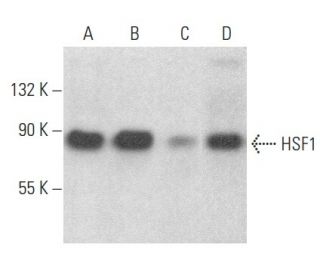
Date published: 2025-9-11

001 800-1338-3838

SCBT Portrait Logo
Seach Input

HSF1 항체 (E-4): sc-17757

4.5(19)
리뷰 쓰기질문하기

데이터 시트
  • HSF1 항체 E-4 는 마우스 monoclonal IgG1 κ HSF1 항체, 51간행물에 인용, 이며 200 µg/ml으로 제공합니다.
  • human origin의 heat shock transcription factor 1 (HSF1)에서 내부에 위치한 219-529 아미노산을 항원으로 사용하였습니다.
  • HSF1 항체 (E-4)는 WB, IP, IF, IHC(P) and ELISA으로 mouse, rat and human유래의 HSF1 를 감지하는 데에 추천한다.
  • 항-HSF1 항체(E-4)는 IP용 agarose와 결합되어 이용 가능하며, WB, IHC(P), ELISA용 HRP와 결합되어 이용 가능하며, IF, IHC(P), FCM용 phycoerythrin 또는 FITC와 결합되어 이용 가능합니다.
  • WB (RGB), IF, IHC(P) 와FCM, RGB fluorescent imaging systems, such as iBright™ FL1000, FluorChem™, Typhoon, Azure and other comparable systems에 사용가능한 Alexa Fluor® 488, Alexa Fluor® 546, Alexa Fluor® 594 or Alexa Fluor® 647결합제품도 있습니다.
  • WB (NIR), IF와FCM,Near-Infrared (NIR) detection systems, such as LI-COR®Odyssey®, iBright™ FL1000, FluorChem™, Typhoon, Azure and other comparable systems에 사용가능한 Alexa Fluor® 680 or Alexa Fluor® 790 결합제품도 있습니다.
  • Gel Supershift와 ChIP 에 사용하는 TransCruz시약(sc-17757 X, 200 µg/0.1 ml)이 있습니다.
  • HSF1 (E-4): sc-17757무료10 µg 샘플을 신청하려면 기술지원팀 (또는 로컬 디스트리뷰터)에 연락주세요.
  • m-IgGκ BP-HRP 는 HSF1 항체 (E-4) 가 WB and IHC(P) 에 응용될 때 우선으로 사용하는 이차 항체 검사 시약입니다. 이 시약은HSF1 항체인 (E-4)와 세트로 제공드립니다。 (아래의 주문 정보를 참고해 주시길 바랍니다)。기타m-IgGκ BP 커플링제는,작은쥐 IgG 결합 단백질완정한 리스트를 참고 해 주시길 바랍니다.

빠른 링크

더보기

HSF1 항체(E-4)는 마우스, 쥐, 인간에서 유래된 HSF1을 웨스턴 블롯(WB), 면역침전(IP), 면역형광(IF), 면역조직화학(IHC), 효소결합 면역흡착 분석법(ELISA)을 통해 검출하는 마우스 단일클론 IgG1 카파 경쇄 항체입니다. HSF1 단일 클론 항체(E-4)는 비접합 형태와 아가로스, 호스래디시 퍼옥시다아제, 피코에리트린, 플루오레세인 이소티오시아네이트, 다중 알렉사 플루오르® 접합체를 포함한 다양한 접합체 형태로 제공됩니다. HSF1은 세포를 손상으로부터 보호하는 데 도움이 되는 열 충격 단백질의 발현을 조절함으로써 스트레스, 특히 열 및 화학적 스트레스에 대한 세포 반응에 중요한 역할을 합니다. 이러한 조절은 세포의 항상성을 유지하고 불리한 조건에서 세포 생존을 촉진하는 데 필수적입니다. HSF1은 일반적으로 단량체로 존재하지만, 활성화되면 핵으로 이동하는 삼량체를 형성하여 HSF1이 표적 유전자의 프로모터 영역에 있는 열 충격 요소에 결합하여 전사를 촉진합니다. HSF1은 에스트로겐에 의해 상향 조절되어 mRNA 및 단백질 수준에서 활성을 향상시키며, 이는 세포 스트레스 반응의 중요성과 스트레스 및 염증과 관련된 질병에 대한 잠재적 영향을 더욱 강조합니다.

연구용으로만 사용가능합니다. 진단이나 치료용으로 사용불가합니다.

Alexa Fluor®는 미국 오리건주 Molecular Probes Inc.의 상표입니다.

LI-COR®와 Odyssey®는 LI-COR Biosciences의 등록 상표입니다.

HSF1 항체 (E-4) 참고문헌:

  1. 진핵생물에서 열충격 유전자의 전사적 활성화.  |  Tanguay, RM. 1988. Biochem Cell Biol. 66: 584-93. PMID: 3048332
  2. 악성 변형에서 HSF1 단백질의 역할.  |  Šimončík, O., et al. 2018. Klin Onkol. 31: 55-62. PMID: 31023025
  3. 종양 미세환경에서 HSF1과 샤페론 네트워크의 역할.  |  Grunberg, N., et al. 2020. Adv Exp Med Biol. 1243: 101-111. PMID: 32297214
  4. HSF1은 간세포암에서 APOJ/STAT3 매개 PD-L1 발현을 조절함으로써 면역치료 반응에 관여한다.  |  Cheng, H., et al. 2023. Cancer Biol Ther. 24: 1-9. PMID: 36482717
  5. 열 충격 인자 1(HSF1)은 표준 열 충격 반응과 관계없이 c-MYC 매개 전사를 강화합니다.  |  Xu, M., et al. 2023. Cell Rep. 42: 112557. PMID: 37224019
  6. 열 충격 유발 자극에 대한 암세포의 전사적 반응은 강력한 HSF1 결합의 증폭을 포함합니다.  |  Dastidar, SG., et al. 2023. Nat Commun. 14: 7420. PMID: 37973875
  7. 열충격 전사인자의 에스트로겐 의존적 발현: 열충격 단백질의 자궁 합성에 대한 시사점.  |  Yang, X., et al. 1995. J Steroid Biochem Mol Biol. 52: 415-9. PMID: 7748806
  8. 주사력 현미경을 이용한 고리형 DNA 복합체에서 열충격 전사인자 2 화학량론 측정.  |  Wyman, C., et al. 1995. EMBO J. 14: 117-23. PMID: 7828583
  9. 마우스 배아 발생 중 열충격 인자 2의 기능 및 조절.  |  Rallu, M., et al. 1997. Proc Natl Acad Sci U S A. 94: 2392-7. PMID: 9122205
  10. 열충격 반응 및 단백질 분해: 유비퀴틴-프로테아좀 경로에 의한 HSF2의 조절.  |  Mathew, A., et al. 1998. Mol Cell Biol. 18: 5091-8. PMID: 9710593
  11. 프로테아좀 억제는 열충격 인자 계열의 모든 구성원의 활성화로 이어집니다.  |  Kawazoe, Y., et al. 1998. Eur J Biochem. 255: 356-62. PMID: 9716376
  12. 글리코겐 합성 효소 키나제 3베타와 세포 외 신호 조절 키나제는 열충격 후 전사 활성 과립의 소멸을 촉진하여 열충격 전사인자 1을 비활성화합니다.  |  He, B., et al. 1998. Mol Cell Biol. 18: 6624-33. PMID: 9774677

주문정보

제품명카탈로그 번호 단위가격수량관심품목

HSF1 항체 (E-4)

sc-17757
200 µg/ml
RMB2377.00

HSF1 (E-4): m-IgGκ BP-HRP 번들

sc-551063
200 µg Ab; 40 µg BP
RMB2662.00

HSF1 항체 (E-4) X

sc-17757 X
200 µg/0.1 ml
RMB2377.00

HSF1 항체 (E-4) AC

sc-17757 AC
500 µg/ml, 25% agarose
RMB3129.00

HSF1 항체 (E-4) HRP

sc-17757 HRP
200 µg/ml
RMB2377.00

HSF1 항체 (E-4) FITC

sc-17757 FITC
200 µg/ml
RMB2482.00

HSF1 항체 (E-4) PE

sc-17757 PE
200 µg/ml
RMB2580.00

HSF1 항체 (E-4) Alexa Fluor® 488

sc-17757 AF488
200 µg/ml
RMB2685.00

HSF1 항체 (E-4) Alexa Fluor® 546

sc-17757 AF546
200 µg/ml
RMB2685.00

HSF1 항체 (E-4) Alexa Fluor® 594

sc-17757 AF594
200 µg/ml
RMB2685.00

HSF1 항체 (E-4) Alexa Fluor® 647

sc-17757 AF647
200 µg/ml
RMB2685.00

HSF1 항체 (E-4) Alexa Fluor® 680

sc-17757 AF680
200 µg/ml
RMB2685.00

HSF1 항체 (E-4) Alexa Fluor® 790

sc-17757 AF790
200 µg/ml
RMB2685.00

Hello, I am a research technician at the University of Bristol, UK. I am interested in your HSF1 (E-4) antibody. Do you have any unconjugated version of this antibody? Can I have a free sample to try it to my cell line. Many thanks.

Asked by: Andriana
Thank you for your question. Please contact Technical Service for information on how to obtain a sample.
Answered by: SCBT Heidelberg Support
Date published: 2018-12-14

I used 3ug of HSF1 (E-4) antibody for doing ChIP, but did not get much enrichment. Is there any recommended concentration of HSF1 antibody for ChIP that I should have used?

Asked by: S_G_
Thank you for your inquiry. For the IP step in ChIP we recommend using 100–500 µg supernatant and 0.1-1 µl TransCruz reagent (0.2–2 µg antibody). The complete protocol for ChIP can be found in below link. If you have any further questions, please call our Technical Service team toll-free at (800) 457-3801 option 2, e-mail <[email protected]>, or use the live chat on our website. Best of luck with your research! https://www.scbt.com/scbt/resources/protocols/chromatin-immunoprecipitation-assays
Answered by: Technical Support 14
Date published: 2017-07-10

What is the recommended secondary reagent to be used with this mouse monoclonal primary?

Asked by: DefinitelyNotMatt
Thank you for your question. We recommend using one our exclusive Mouse IgG Binding Proteins as a secondary detection reagent. A complete list of available binding proteins is available on our website here: https://www.scbt.com/scbt/browse/support-products-mouse-igg-binding-proteins/_/N-ecrety Please contact Technical Service if you have further questions concerning this product.
Answered by: Technical Support 12
Date published: 2017-01-13

Can I use this mouse monoclonal antibody for immunohistochemical staining of mouse tissue? Or even double or triple staining on the same samples?

Asked by: DefinitelyNotMatt
Thank you for your question. Yes. We recommend using a directly conjugated mouse monoclonal primary antibody. This antibody is currently available conjugated to phycoerythrin (sc-17757 PE), fluorescein (sc-17757 FITC), Alexa Fluor® 488 (sc-17757 AF488) or Alexa Fluor® 647 (sc-17757 AF647). Please contact Technical Service if you have further questions concerning this product.
Answered by: Technical Support 12
Date published: 2017-01-13
  • y_2025, m_9, d_11, h_4CST
  • bvseo_bulk, prod_bvqa, vn_bulk_3.0.42
  • cp_1, bvpage1
  • co_hasquestionsanswers, tq_4
  • loc_ko_KR, sid_17757, prod, sort_[SortEntry(order=LAST_APPROVED_ANSWER_SUBMISSION_TIME, direction=DESCENDING)]
  • clientName_scbt
  • bvseo_sdk, java_sdk, bvseo-4.0.0
  • CLOUD, getContent, 99ms
  • QUESTIONS, PRODUCT
Rated 5 out of 5 by from works goodworks good for my sample. cell line over-expressed with HSF1. 1:1000, clear background. but not sure for detecting endogenous level.
Date published: 2022-01-25
Rated 1 out of 5 by from Not good for WB using human whole cell lysate.This antibody is not good for WB using human K562 whole cell lysate at 1:500 dilution.
Date published: 2021-08-05
Rated 2 out of 5 by from Not working for my samplesI’ve tried this antibody on sub cellular fractionated PBMCs Lysates at 1:100 dilution but the signal was very low
Date published: 2020-09-02
Rated 3 out of 5 by from works okayfaint signals were observed - samples were human cells
Date published: 2017-11-21
Rated 5 out of 5 by from specifc signaWe got specific bands at 90kda with diution 1:800 by WB.HepG2 cell lysates 60ug.I don't recommend loading than 80ug, we have tried this amout ,it cause very high background,also I recommend use the secondary antibody sc-516102.
Date published: 2017-09-16
Rated 5 out of 5 by from 功能很出色the antibody is very good,economical and practical
Date published: 2017-09-12
Rated 4 out of 5 by from acceptable antibody for immunofluorescenceDilution 1:200 for IF on PFA fixed U2OS cells gives week signal (compared to polyclonal rabbit Ab). 1:50 should be tested.
Date published: 2017-08-08
Rated 5 out of 5 by from specific binding target proteinI use HSF1antibody in our WB assay.The antibody is very full bind the target protein,specific and 1:500 is suitable but for the loading amount I recommend controled at 40 to 70 ug lysates.If more than this range,it may affect the specificity and singal intensity.
Date published: 2017-07-15
  • y_2025, m_9, d_11, h_4
  • bvseo_bulk, prod_bvrr, vn_bulk_3.0.42
  • cp_1, bvpage1
  • co_hasreviews, tv_0, tr_19
  • loc_ko_KR, sid_17757, prod, sort_[SortEntry(order=SUBMISSION_TIME, direction=DESCENDING)]
  • clientName_scbt
  • bvseo_sdk, java_sdk, bvseo-4.0.0
  • CLOUD, getReviews, 10ms
  • REVIEWS, PRODUCT
HSF1 항체 (E-4) is rated 4.5 out of 5 by 19.
  • y_2025, m_9, d_11, h_4
  • bvseo_bulk, prod_bvrr, vn_bulk_3.0.42
  • cp_1, bvpage1
  • co_hasreviews, tv_0, tr_19
  • loc_ko_KR, sid_17757, prod, sort_[SortEntry(order=SUBMISSION_TIME, direction=DESCENDING)]
  • clientName_scbt
  • bvseo_sdk, java_sdk, bvseo-4.0.0
  • CLOUD, getAggregateRating, 118ms
  • REVIEWS, PRODUCT