")
Dok-1 항체 (A-3): sc-6929
- Dok-1 항체 A-3 는 마우스 monoclonal IgG1 κ Dok-1 항체, 19간행물에 인용, 이며 200 µg/ml으로 제공합니다.
- 아미노 산1-276 mapping at the N-terminus of Dok-1 of mouse 의하고 기원한다
- Dok-1 항체 (A-3)는 WB, IP, IF and IHC(P)으로 mouse, rat and human유래의 Dok-1 를 감지하는 데에 추천한다.
- 항-Dok-1 항체(A-3)는 IP용 agarose와 결합되어 이용 가능하며, WB, IHC(P), ELISA용 HRP와 결합되어 이용 가능하며, IF, IHC(P), FCM용 phycoerythrin 또는 FITC와 결합되어 이용 가능합니다.
- WB (RGB), IF, IHC(P) 와FCM, RGB fluorescent imaging systems, such as iBright™ FL1000, FluorChem™, Typhoon, Azure and other comparable systems에 사용가능한 Alexa Fluor® 488, Alexa Fluor® 546, Alexa Fluor® 594 or Alexa Fluor® 647결합제품도 있습니다.
- WB (NIR), IF와FCM,Near-Infrared (NIR) detection systems, such as LI-COR®Odyssey®, iBright™ FL1000, FluorChem™, Typhoon, Azure and other comparable systems에 사용가능한 Alexa Fluor® 680 or Alexa Fluor® 790 결합제품도 있습니다.
- m-IgG Fc BP-HRP, 1 BP-HRP">m-IgG1 BP-HRP 및 m-IgGκ BP-HRP는 Dok-1 항체(A-3) WB 및 IHC(P) 애플리케이션용입니다. 이 시약은 이제 Dok-1 항체(A-3)와 함께 번들로 제공됩니다(아래 주문 정보 참조).

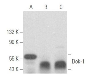
Dok-1 항체(A-3)는 마우스, 랫트, 인간 유래의 Dok-1 단백질을 웨스턴 블롯(WB), 면역침전(IP), 면역형광(IF), 면역조직화학(IHC)을 통해 검출하는 마우스 단일클론 IgG1 카파 경쇄 항체입니다. Dok-1 (A-3) 항체는 비접합 형태와 아가로스, 과산화효소(HRP), 피코에리트린(PE), 플루오레세인 이소티오시아네이트(FITC), 다중 알렉사 플루오르® 접합체를 포함한 다양한 접합체 형태로 제공됩니다. Dok-1은 다양한 신호 전달 분자와 상호 작용하는 도킹 단백질로서 작용함으로써 세포 신호 전달 경로, 특히 세포 성장과 분화 조절에 중요한 역할을 합니다. 티로신 인산화 시 Ras GTPase 활성화 단백질(Ras GAP)과 결합하는 Dok-1의 능력은 매우 중요합니다. 이 결합은 세포 증식과 생존에 중요한 역할을 하는 Ras의 활동을 조절하는 데 도움이 되기 때문입니다. 또한, Dok-1은 혈소판 유래 성장 인자(PDGF), 혈관 내피 성장 인자(VEGF), 인슐린, 인슐린 유사 성장 인자(IGF)를 포함한 여러 성장 인자에 의해 활성화되는 신호 전달 경로에 관여하고 있으며, 이는 Dok-1이 정상적인 생리학 및 만성 골수성 백혈병과 같은 질병 상태 모두에서 중요하다는 것을 강조합니다. 또한, Dok-1은 IRS-1 및 Cas와 같은 다른 티로신 키나아제 기질과 함께 다수의 티로신 잔기와 추정 SH2 결합 부위를 포함하고 있으며, 이는 신호 전달에서 Dok-1의 기능에 필수적입니다.
Alexa Fluor®는 미국 오리건주 Molecular Probes Inc.의 상표입니다.
LI-COR®와 Odyssey®는 LI-COR Biosciences의 등록 상표입니다.
Dok-1 항체 (A-3) 참고문헌:
- Dok-1과 Dok-2 결핍은 파골 세포의 활성화를 통해 골감소증을 유발합니다. | Kawamata, A., et al. 2011. J Cell Physiol. 226: 3087-93. PMID: 21732353
- Dok-1은 혈소판 인테그린 αIIbβ3 외부 유입 신호를 부정적으로 조절하고 생쥐의 혈전증을 억제합니다. | Niki, M., et al. 2016. Thromb Haemost. 115: 969-78. PMID: 26790499
- Dok-1과 Dok-2는 기억 CD8+ T 세포의 형성을 조절합니다. | Laroche-Lefebvre, C., et al. 2016. J Immunol. 197: 3618-3627. PMID: 27664281
- Dok-1은 칼슘 의존성 F-액틴 분해를 억제하여 비만 세포 탈과립화를 부정적으로 조절합니다. | Du, H., et al. 2022. Clin Immunol. 238: 109008. PMID: 35421591
- SH2 도메인이 단백질 티로신 키나아제에 의한 과정적 인산화를 촉진한다는 증거. | Mayer, BJ., et al. 1995. Curr Biol. 5: 296-305. PMID: 7780740
- 혈관 내피 세포 성장 인자는 SH2 도메인을 포함하는 신호 전달 매개체의 티로신 인산화를 촉진합니다. 내피 세포 증식과의 연관성. | Guo, D., et al. 1995. J Biol Chem. 270: 6729-33. PMID: 7896817
- IRS-1 신호 체계. | Myers, MG., et al. 1994. Trends Biochem Sci. 19: 289-93. PMID: 8048169
- 원발성 만성기 만성 골수성 백혈병 농축 혈통 음성 폭발 집단에 구성적으로 존재하는 62킬로달톤 티로신 인산화단백질. | Wisniewski, D., et al. 1994. Leukemia. 8: 688-93. PMID: 8152267
- EGF 및 인슐린 수용체 신호에서 Grb2 관련 도킹 단백질. | Holgado-Madruga, M., et al. 1996. Nature. 379: 560-4. PMID: 8596638
- p62(독): 만성 골수성 백혈병 전구 세포에서 구성적으로 티로신이 인산화되고 GAP와 관련된 단백질입니다. | Carpino, N., et al. 1997. Cell. 88: 197-204. PMID: 9008160
- 도킹 단백질인 Dok으로 Abl 및 rasGAP 관련 62kDa 단백질의 동정. | Yamanashi, Y. and Baltimore, D. 1997. Cell. 88: 205-11. PMID: 9008161
- p56dok-2의 분자 복제 및 특성 분석으로 새로운 RasGAP 결합 단백질 계열 정의. | Di Cristofano, A., et al. 1998. J Biol Chem. 273: 4827-30. PMID: 9478921
주문정보
| 제품명 | 카탈로그 번호 | 단위 | 가격 | 수량 | 관심품목 | |
Dok-1 항체 (A-3) | sc-6929 | 200 µg/ml | CNY2422.00 | |||
Dok-1 (A-3): m-IgG Fc BP-HRP 번들 | sc-528168 | 200 µg Ab; 10 µg BP | CNY2715.00 | |||
Dok-1 (A-3): m-IgGκ BP-HRP 번들 | sc-520478 | 200 µg Ab, 40 µg BP | CNY2715.00 | |||
Dok-1 (A-3): m-IgG1 BP-HRP 번들 | sc-542796 | 200 µg Ab; 20 µg BP | CNY2715.00 | |||
Dok-1 항체 (A-3) AC | sc-6929 AC | 500 µg/ml, 25% agarose | CNY3189.00 | |||
Dok-1 항체 (A-3) HRP | sc-6929 HRP | 200 µg/ml | CNY2422.00 | |||
Dok-1 항체 (A-3) FITC | sc-6929 FITC | 200 µg/ml | CNY2527.00 | |||
Dok-1 항체 (A-3) PE | sc-6929 PE | 200 µg/ml | CNY2625.00 | |||
Dok-1 항체 (A-3) Alexa Fluor® 488 | sc-6929 AF488 | 200 µg/ml | CNY2738.00 | |||
Dok-1 항체 (A-3) Alexa Fluor® 546 | sc-6929 AF546 | 200 µg/ml | CNY2738.00 | |||
Dok-1 항체 (A-3) Alexa Fluor® 594 | sc-6929 AF594 | 200 µg/ml | CNY2738.00 | |||
Dok-1 항체 (A-3) Alexa Fluor® 647 | sc-6929 AF647 | 200 µg/ml | CNY2738.00 | |||
Dok-1 항체 (A-3) Alexa Fluor® 680 | sc-6929 AF680 | 200 µg/ml | CNY2738.00 | |||
Dok-1 항체 (A-3) Alexa Fluor® 790 | sc-6929 AF790 | 200 µg/ml | CNY2738.00 |