")
: sc-130914. 핵 국소화를 보여주는 메탄올 고정 HeLa 세포의 면역 형광 염색.")
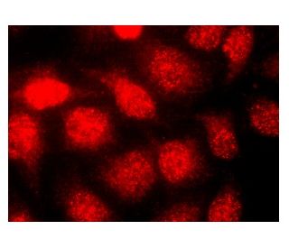
8-oxoG DNA 병변 항체(483.15): sc-130914. 핵 국소화를 보여주는 메탄올 고정 HeLa 세포의 면역 형광 염색.
8-oxoG DNA Lesion 항체 (483.15): sc-130914
- 8-oxoG DNA Lesion 항체 483.15 는 마우스 monoclonal IgM κ 8-oxoG DNA Lesion 항체, 23간행물에 인용, 이며 200 µg/ml으로 제공합니다.
- 쥐 간에서 분리된 DOC 불용성 접합 리본에 대해 제기됨
- IF와 ELISA으로 8-oxoG Lesions 을 검출할것을 권장합니다.
- 현재 8-oxoG DNA Lesion 항체(483.15)에 대해 선호하는 2차 검출 시약의 식별을 아직 완료하지 못했습니다. 이 작업은 진행 중입니다.

빠른 링크
보조 제품
설명
데이터 시트 및 프로토콜
연구 정보
더보기
8-oxoG DNA 병변 항체(483.15)는 8-oxoG DNA 병변 단백질을 검출하는 IgM κ 마우스 단일 클론 8-oxoG DNA 병변 항체(8-oxoG DNA 병변 항체로도 지정됨)입니다. 8-oxoG DNA 병변 항체(483.15)는 비접합 항 8-oxoG DNA 병변 항체로서 사용 가능합니다. DNA(디옥시리보핵산)는 알려진 모든 생물체와 일부 바이러스의 유전 물질입니다. 정보의 장기 저장과 발달 및 기능에 중요한 역할을 하는 DNA는 당 백본과 인산염 그룹을 포함하는 두 개의 긴 평행 뉴클레오티드 폴리머로 구성되어 있으며 에스테르 결합으로 서로 결합되어 이중 나선을 형성합니다. 8-oxoG(8-옥소구아닌)는 산화성 DNA 손상에 관여하는 돌연변이 유발성 병변입니다. 8-oxoG는 DNA 복제 중에 아데닌(A)과 잘못 결합하여 게놈 안정성에 위협을 가할 수 있습니다. 모든 유기체는 8-oxoG를 복구하기 위해 최소 두 가지 유형의 8-oxoG-DNA 글리코실라아제(OGG)를 발현합니다.
연구용으로만 사용가능합니다. 진단이나 치료용으로 사용불가합니다.
Alexa Fluor®는 미국 오리건주 Molecular Probes Inc.의 상표입니다.
LI-COR®와 Odyssey®는 LI-COR Biosciences의 등록 상표입니다.
8-oxoG DNA Lesion 항체 (483.15) 참고문헌:
- 8-옥소구아닌 복구를 위한 여러 DNA 글리코실라제 및 생체 내 잠재적 기능. | Hazra, TK., et al. 2001. Prog Nucleic Acid Res Mol Biol. 68: 193-205. PMID: 11554297
- CHO 세포주에서 8-옥소구아닌 및 Ogg1-절개된 아퓨린 부위의 복구. | Boiteux, S. and le Page, F. 2001. Prog Nucleic Acid Res Mol Biol. 68: 95-105. PMID: 11554315
- 활성산소 과잉 생성에 취약한 노화 OXYS 쥐의 간세포에서 8-옥소구아닌 DNA 병변을 면역 형광으로 검출합니다. | Kemeleva, EA., et al. 2006. Mutat Res. 599: 88-97. PMID: 16574166
- 8-옥소구아닌의 위협에 대처하기 위한 다양한 DNA 복구 전략. | Russo, MT., et al. 2007. Mutat Res. 614: 69-76. PMID: 16769088
- 8-옥소구아닌에서의 전사 돌연변이 유발의 분자적 기초. | Damsma, GE. and Cramer, P. 2009. J Biol Chem. 284: 31658-63. PMID: 19758983
- 전기화학적 검출을 통한 ELISA 또는 고성능 액체 크로마토그래피(HPLC)를 이용한 산화적 DNA 손상 8-옥소구아닌(8-oxoG)의 측정. | Drake, DM., et al. 2019. Methods Mol Biol. 1965: 313-328. PMID: 31069684
- HDAC1은 노화된 뇌와 알츠하이머병에서 OGG1에 의한 산화성 DNA 손상 복구를 조절합니다. | Pao, PC., et al. 2020. Nat Commun. 11: 2484. PMID: 32424276
- 식물 텔로미어에서 8-oxoG의 정량화. | Castillo-González, C., et al. 2022. Int J Mol Sci. 23: PMID: 35563379
- 산화적 DNA 손상 8-OxoG를 종양 발생과 진행에 연결. | Zhao, Y., et al. 2022. Yi Chuan. 44: 466-477. PMID: 35729095
- 종양 발생 및 암 치료에서 8-oxoG 복구 시스템의 역할. | Li, C., et al. 2022. Cells. 11: PMID: 36497058
- 핵 추출물(SMADNE)에서 DNA 결합 단백질의 단일 분자 분석. | Schaich, MA., et al. 2023. Nucleic Acids Res. 51: e39. PMID: 36861323
- 인간 세포에는 두 가지 다른 8-옥소구아닌 복구 효소가 존재하며, 돌연변이를 예방하는 데 잠재적으로 상호 보완적인 역할을 합니다. | Hazra, TK., et al. 1998. Nucleic Acids Res. 26: 5116-22. PMID: 9801308
주문정보
| 제품명 | 카탈로그 번호 | 단위 | 가격 | 수량 | 관심품목 | |
8-oxoG DNA Lesion 항체 (483.15) | sc-130914 | 200 µg/ml | CNY2422.00 |