")
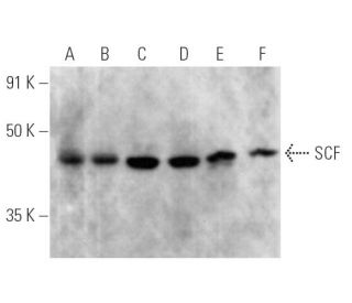
SCF 抗体 (G-3): sc-13126. MG-63 (A), NCI-H1299 (B), NIH/3T3 (C), Neuro-2A (D), C6 (E) 和 PC-12 (F) 全细胞裂解液中 SCF 表达的 Western 印迹分析.
SCF 抗体 (G-3): sc-13126
- SCF 抗体 G-3 是小鼠单克隆 IgG2b κ,SCF抗体, 在53篇文献中引用,规格为200 µg/ml
- 免疫human物种的stem cell factor (SCF)的氨基酸26-214
- SCF 抗体 (G-3) 推荐用于 WB, IP, IF, IHC(P) 和 ELISA,检测mouse, rat 和human 来源的 SCF
- 抗SCF抗体(G-3)可与琼脂糖结合用于IP;与HRP结合用于WB、IHC(P)和ELISA;与藻红蛋白或FITC结合用于IF、IHC(P)和FCM
- 还可偶联Alexa Fluor® 488, Alexa Fluor® 546, Alexa Fluor® 594 和 Alexa Fluor® 647,用于WB (RGB), IF, IHC(P) 和 FCM, 以及用于RGB荧光成像系统,例如iBright™ FL1000, FluorChem™, Typhoon, Azure和其他类似的系统
- 还可偶联Alexa Fluor® 680 和 Alexa Fluor® 790, 用于WB (NIR), IF 和 FCM; 以及用于近红外(NIR)检测系统,如LI-COR®/Odyssey®, iBright™ FL1000, FluorChem™, Typhoon, Azure和类似系统
- 2b BP-HRP">m-IgG2b BP-HRP和m-IgGκ BP-HRP是SCF Antibody (G-3) 适用于 WB 和 IHC(P) 应用。 的首选辅助检测试剂。这些试剂现与SCF Antibody (G-3) 打包提供(请参阅下面的订购信息)。

快捷链接
相关产品
描述
基因信息
蛋白序列
说明书与实验方案
研究信息
関連項目
SCF抗体(G-3)是一种小鼠单克隆IgG2b kappa轻链抗体,可通过WB、IP、IF、IHCP和ELISA检测小鼠、大鼠和人源SCF蛋白。SCF抗体(G-3)有非结合型和多种结合型,包括琼脂糖、HRP、PE、FITC和多种Alexa Fluor®结合型。SCF作为跨膜酪氨酸激酶受体原癌基因c-Kit的配体,在多种细胞过程中发挥着至关重要的作用。SCF是一种多效细胞因子,在人类和小鼠体内分别由248个和220个氨基酸组成,以两种可替代的剪接形式存在。较大的SCF形式主要在成纤维细胞、大脑和胸腺中表达,而较小的变异形式则存在于脾脏、睾丸、胎盘和小脑等组织中。SCF对于生殖细胞、造血祖细胞和黑色素细胞前体的发育至关重要,因为SCF能够刺激成熟和未成熟肥大细胞的增殖和成熟。SCF与c-Kit的相互作用对于包括造血和色素沉着在内的各种生物过程至关重要,这使得SCF单克隆抗体(G-3)成为发育生物学和癌症研究领域的重要研究工具。
仅限研究使用。不适用于诊断和治疗用途。
Alexa Fluor® 是Molecular Probes Inc., OR., USA的商标
LI-COR®和 Odyssey® 是LI-COR Biosciences的注册商标
SCF 抗体 (G-3) 参考文献:
- KL-1 和 KL-2 两种细胞缔合作用形式的差异表达和处理:KL-1 和 KL-2。 | Huang, EJ., et al. 1992. Mol Biol Cell. 3: 349-62. PMID: 1378327
- 小鼠基质细胞选择性地表达膜结合型和分泌型的人类同源钢基因产物--干细胞因子,从而在长期骨髓培养中支持人类造血。 | Toksoz, D., et al. 1992. Proc Natl Acad Sci U S A. 89: 7350-4. PMID: 1380155
- 膜锚定生长因子的裂解涉及通过共同机制调节的不同蛋白酶活性。 | Pandiella, A., et al. 1992. J Biol Chem. 267: 24028-33. PMID: 1385433
- 肥大细胞生长因子位于小鼠第 10 号染色体的钢基因座附近,在一些钢等位基因中被删除。 | Copeland, NG., et al. 1990. Cell. 63: 175-83. PMID: 1698554
- 大鼠和人类干细胞因子 DNA 的初级结构和功能表达。 | Martin, FH., et al. 1990. Cell. 63: 203-11. PMID: 2208279
- 干细胞因子在胎盘微环境中的作用。 | Khodadi, E., et al. 2016. Cell Tissue Res. 366: 523-531. PMID: 27234501
- 促红细胞生成素、干细胞因子和癌细胞迁移。 | Vazquez-Mellado, MJ., et al. 2017. Vitam Horm. 105: 273-296. PMID: 28629522
- 骨髓脂肪细胞通过分泌 SCF 促进干细胞再生和造血。 | Zhou, BO., et al. 2017. Nat Cell Biol. 19: 891-903. PMID: 28714970
- 干细胞因子:骨髓脂肪细胞和造血细胞之间的桥梁。 | Li, Z. and MacDougald, OA. 2019. Haematologica. 104: 1689-1691. PMID: 31473604
- 抑制可溶性干细胞因子可促进肠粘膜修复。 | Garcia-Hernandez, V., et al. 2023. Inflamm Bowel Dis. 29: 1133-1144. PMID: 36688460
- 干细胞因子和cKIT调节缺氧状态下的内皮细胞糖酵解。 | Jeong, H., et al. 2024. Cardiovasc Res. 120: 745-755. PMID: 38507654
- 在有 SCF 的情况下培养单核细胞不会制造肥大细胞。循环肥大细胞祖细胞的特征为 c-kit+, CD34+, Ly-, CD14-, CD17-, 集落形成细胞。 | Agis, H., et al. 1993. J Immunol. 151: 4221-7. PMID: 7691941
订购信息
| 产品名称 | 产品编号 | 规格 | 价格 | 数量 | 收藏夹 | |
SCF 抗体 (G-3) | sc-13126 | 200 µg/ml | CNY2422.00 | |||
SCF (G-3): m-IgGκ BP-HRP 套装 | sc-520598 | 200 µg Ab, 40 µg BP | CNY2715.00 | |||
SCF (G-3): m-IgG2b BP-HRP 套装 | sc-548841 | 200 µg Ab; 10 µg BP | CNY2715.00 | |||
SCF 抗体 (G-3) AC | sc-13126 AC | 500 µg/ml, 25% agarose | CNY3189.00 | |||
SCF 抗体 (G-3) HRP | sc-13126 HRP | 200 µg/ml | CNY2422.00 | |||
SCF 抗体 (G-3) FITC | sc-13126 FITC | 200 µg/ml | CNY2527.00 | |||
SCF 抗体 (G-3) PE | sc-13126 PE | 200 µg/ml | CNY2625.00 | |||
SCF 抗体 (G-3) Alexa Fluor® 488 | sc-13126 AF488 | 200 µg/ml | CNY2738.00 | |||
SCF 抗体 (G-3) Alexa Fluor® 546 | sc-13126 AF546 | 200 µg/ml | CNY2738.00 | |||
SCF 抗体 (G-3) Alexa Fluor® 594 | sc-13126 AF594 | 200 µg/ml | CNY2738.00 | |||
SCF 抗体 (G-3) Alexa Fluor® 647 | sc-13126 AF647 | 200 µg/ml | CNY2738.00 | |||
SCF 抗体 (G-3) Alexa Fluor® 680 | sc-13126 AF680 | 200 µg/ml | CNY2738.00 | |||
SCF 抗体 (G-3) Alexa Fluor® 790 | sc-13126 AF790 | 200 µg/ml | CNY2738.00 |