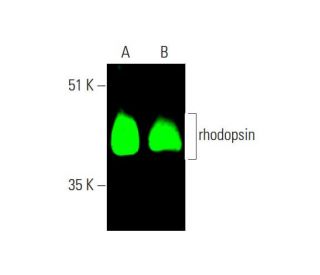
Date published: 2025-9-7

021-6093-6350

SCBT Portrait Logo
Seach Input

rhodopsin 抗体 (1D4): sc-57432

4.4(12)
寫評論提問

说明书
  • rhodopsin 抗体 1D4 是小鼠单克隆 IgG1 κ,rhodopsin抗体, 在77篇文献中引用,规格为200 µg/ml
  • 针对全长rhodopsin,bovine起源
  • rhodopsin 抗体 (1D4) 推荐用于 WB, IP, IF, IHC(P) 和 ELISA,检测mouse, rat 和human 来源的 rhodopsin; 也和以下物种反应,包括: bovine
  • 抗rhodopsin抗体(1D4)可与琼脂糖结合用于IP;与HRP结合用于WB、IHC(P)和ELISA;与藻红蛋白FITC结合用于IF、IHC(P)和FCM
  • 还可偶联Alexa Fluor® 488, Alexa Fluor® 546, Alexa Fluor® 594Alexa Fluor® 647,用于WB (RGB), IF, IHC(P) 和 FCM, 以及用于RGB荧光成像系统,例如iBright™ FL1000, FluorChem™, Typhoon, Azure和其他类似的系统
  • 还可偶联Alexa Fluor® 680Alexa Fluor® 790, 用于WB (NIR), IF 和 FCM; 以及用于近红外(NIR)检测系统,如LI-COR®/Odyssey®, iBright™ FL1000, FluorChem™, Typhoon, Azure和类似系统
  • 关于如何获取rhodopsin (1D4): sc-57432免费10 µg小样,联系我们技术服务部门 (或者您当地的代理商)了解详情。
  • m-IgG Fc BP-HRP, m-IgG1 BP-HRPm-IgGκ BP-HRP 是 rhodopsin 抗体 (1D4) 在 WB and IHC(P) 应用中的首选二抗检测试剂。这些试剂现与rhodopsin 抗体 (1D4) 组合成套装提供。 (请参阅下面的订购信息)。

快捷链接

関連項目

rhodopsin 抗体(1D4)是一种小鼠单克隆 IgG1 kappa 轻链抗体,可通过西部印迹(WB)、免疫沉淀(IP)、免疫荧光(IF)、免疫组织化学和酶联免疫吸附试验(ELISA)检测小鼠、大鼠和人源的 rhodopsin 蛋白。抗泪腺素抗体 (1D4) 有非共轭型和多种共轭型,包括琼脂糖、辣根过氧化物酶 (HRP)、藻红蛋白 (PE)、异硫氰酸荧光素 (FITC) 和多种 Alexa Fluor® 共轭物。视紫红质是 G 蛋白偶联受体(GPCR)视蛋白亚家族的成员,在视觉光传导途径中发挥着至关重要的作用,使生物体能够感知光线。视网膜上的视紫红质位于杆状感光细胞的外节,对弱光条件下的视觉至关重要。这种蛋白质由共价结合的发色团--11-顺式视网膜色素组成,在吸收光子时发生异构化,引发一系列构象变化,从而激活 G 蛋白,启动负责视觉感知的信号级联。这一过程能够在昏暗环境中检测光线,对夜视和整体视觉敏锐度至关重要。视网膜色素变性是一种退行性眼病,可导致严重的视力丧失,因此视网膜色素基因突变可导致各种形式的视网膜色素变性,这凸显了视网膜色素在维持视网膜健康和功能方面的重要性。

仅限研究使用。不适用于诊断和治疗用途。

Alexa Fluor® 是Molecular Probes Inc., OR., USA的商标

LI-COR®和 Odyssey® 是LI-COR Biosciences的注册商标

rhodopsin 抗体 (1D4) 参考文献:

  1. 视紫红质的光异构化。  |  Kandori, H., et al. 2001. Biochemistry (Mosc). 66: 1197-209. PMID: 11743865
  2. 视紫红质介导的视网膜色素变性。  |  Malanson, KM. and Lem, J. 2009. Prog Mol Biol Transl Sci. 88: 1-31. PMID: 20374723
  3. 视紫红质对磷脂的扰乱。  |  Ernst, OP. and Menon, AK. 2015. Photochem Photobiol Sci. 14: 1922-31. PMID: 26179029
  4. 视紫红质低聚化和聚集。  |  Park, PS. 2019. J Membr Biol. 252: 413-423. PMID: 31286171
  5. 视觉的光触发环核苷酸级联中的信息流。  |  Fung, BK., et al. 1981. Proc Natl Acad Sci U S A. 78: 152-6. PMID: 6264430
  6. 牛视网膜红蛋白的结构。  |  Hargrave, PA., et al. 1983. Biophys Struct Mech. 9: 235-44. PMID: 6342691
  7. G 蛋白疾病为开启开关提供了一个模型。  |  Iiri, T., et al. 1998. Nature. 394: 35-8. PMID: 9665125
  8. 视紫红质的结构。  |  Schertler, GF. 1998. Eye (Lond). 12 (Pt 3b): 504-10. PMID: 9775210

订购信息

产品名称产品编号规格价格数量收藏夹

rhodopsin 抗体 (1D4)

sc-57432
200 µg/ml
RMB2377.00

rhodopsin (1D4): m-IgG Fc BP-HRP 套装

sc-528623
200 µg Ab; 10 µg BP
RMB2662.00

rhodopsin (1D4): m-IgGκ BP-HRP 套装

sc-521049
200 µg Ab, 40 µg BP
RMB2662.00

rhodopsin (1D4): m-IgG1 BP-HRP 套装

sc-543035
200 µg Ab; 20 µg BP
RMB2662.00

rhodopsin 抗体 (1D4) AC

sc-57432 AC
500 µg/ml, 25% agarose
RMB3129.00

rhodopsin 抗体 (1D4) HRP

sc-57432 HRP
200 µg/ml
RMB2377.00

rhodopsin 抗体 (1D4) FITC

sc-57432 FITC
200 µg/ml
RMB2482.00

rhodopsin 抗体 (1D4) PE

sc-57432 PE
200 µg/ml
RMB2580.00

rhodopsin 抗体 (1D4) Alexa Fluor® 488

sc-57432 AF488
200 µg/ml
RMB2685.00

rhodopsin 抗体 (1D4) Alexa Fluor® 546

sc-57432 AF546
200 µg/ml
RMB2685.00

rhodopsin 抗体 (1D4) Alexa Fluor® 594

sc-57432 AF594
200 µg/ml
RMB2685.00

rhodopsin 抗体 (1D4) Alexa Fluor® 647

sc-57432 AF647
200 µg/ml
RMB2685.00

rhodopsin 抗体 (1D4) Alexa Fluor® 680

sc-57432 AF680
200 µg/ml
RMB2685.00

rhodopsin 抗体 (1D4) Alexa Fluor® 790

sc-57432 AF790
200 µg/ml
RMB2685.00

Hi could you send our lab a sample antibody of Rhodopsin 1D4

Asked by: Wenjing Wu
Thank you for contacting Santa Cruz Biotechnology . To receive details about our sample program please email [email protected]
Answered by: BlakeJ
Date published: 2024-06-27

Would this antibody recognize the 1D4 tag in fusion protein in western blot?

Asked by: rogomca
Thank you for your question. This is a mouse monoclonal antibody that is recommended for detection of the protein rhodopsin. The clone name for this antibody is “1D4”, which is also known as the “1D4” epitope of rhodopsin. We have not tested this antibody for detection of any 1D4 fusion proteins.
Answered by: Technical Support
Date published: 2022-09-14

Can this mouse monoclonal antibody be used in IF or IHC on mouse tissue without background or non-specific staining issues?

Asked by: Anonymous
Thank you for your question. Yes, it can be used successfully for this application. We recommend using a directly conjugated mouse monoclonal primary antibody. This mouse monoclonal antibody rhodopsin (1D4): sc-57432 is currently available conjugated to HRP, PE, FITC, AF488 and AF647.
Answered by: Technical Support 10
Date published: 2017-01-09
  • y_2025, m_9, d_7, h_4CST
  • bvseo_bulk, prod_bvqa, vn_bulk_3.0.42
  • cp_1, bvpage1
  • co_hasquestionsanswers, tq_3
  • loc_zh_CN, sid_57432, prod, sort_[SortEntry(order=LAST_APPROVED_ANSWER_SUBMISSION_TIME, direction=DESCENDING)]
  • clientName_scbt
  • bvseo_sdk, java_sdk, bvseo-4.0.0
  • CLOUD, getContent, 149ms
  • QUESTIONS, PRODUCT
Rated 5 out of 5 by from Buen resultadoEl patrón de expresión del anticuerpo fue el esperado en las dos especies de peces utilizadas.
Date published: 2021-03-25
Rated 4 out of 5 by from Visualizing Endogenous Rhodopsin in RPE CellsUseful antibody for IF labeling of endogenous rhodopsin in RPE cells. Cells permeabilized for 30min in PBStween then blocked in 5%BSA. Antibody used at 1:200 overnight at 4 degrees.
Date published: 2020-02-14
Rated 4 out of 5 by from Antibody for IFI used this antibody on OCT embedded frozen retinal sections (mouse). Blocked for 1 hour with 10% goat serum buffer. Incubated with primary antibody overnight at the a concentration of 1:100. Incubated with secondary antibody (alexa fluor 568 anti-mouse) at a concentration of 1:1000. Staining was fairly robust but not completely consistent between all samples.
Date published: 2018-03-21
Rated 2 out of 5 by from Poor specificity in normal canine WBAntibody was used for WB with normal canine tissue. Very low specificity.
Date published: 2017-08-18
Rated 5 out of 5 by from Great reactivity.This antibody will give you strong and specific signals in your blotting and staining. It even cross-reacts with additional species. It has been published with over 15 times.
Date published: 2017-01-11
Rated 5 out of 5 by from Outstanding!!Great product for both western blot and ihc cell staining applications
Date published: 2017-01-05
Rated 4 out of 5 by from DecentI bought this antibody for WB of rhodopsin in Y79 cells. So far it works as advertised.
Date published: 2016-12-12
Rated 4 out of 5 by from Good specificity and signal strength withGood specificity and signal strength with some background in mouse eye and rat eye tissue extracts. -SCBT QC
Date published: 2015-06-01
  • y_2025, m_9, d_7, h_4
  • bvseo_bulk, prod_bvrr, vn_bulk_3.0.42
  • cp_1, bvpage1
  • co_hasreviews, tv_0, tr_12
  • loc_zh_CN, sid_57432, prod, sort_[SortEntry(order=SUBMISSION_TIME, direction=DESCENDING)]
  • clientName_scbt
  • bvseo_sdk, java_sdk, bvseo-4.0.0
  • CLOUD, getReviews, 18ms
  • REVIEWS, PRODUCT
rhodopsin 抗体 (1D4) is rated 4.4 out of 5 by 12.
  • y_2025, m_9, d_7, h_4
  • bvseo_bulk, prod_bvrr, vn_bulk_3.0.42
  • cp_1, bvpage1
  • co_hasreviews, tv_0, tr_12
  • loc_zh_CN, sid_57432, prod, sort_[SortEntry(order=SUBMISSION_TIME, direction=DESCENDING)]
  • clientName_scbt
  • bvseo_sdk, java_sdk, bvseo-4.0.0
  • CLOUD, getAggregateRating, 108ms
  • REVIEWS, PRODUCT