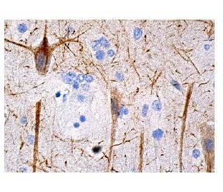
Date published: 2025-9-8

021-6093-6350

SCBT Portrait Logo
Seach Input

NF-H 抗体 (A-12): sc-133165

5.0(3)
寫評論提問

说明书
  • NF-H抗体(A-12)是小鼠单克隆IgG1 κ, 在2篇文献中引用,规格为200 µg/ml
  • 针对氨基酸1-100,在NF-H的human起源的N-terminus处进行映射
  • 推荐用于 mouse, rat 和 human 来源的NF-H WB, IP, IF, IHC(P) 和 ELISA检测
  • m-IgG Fc BP-HRPm-IgG1 BP-HRP 是 NF-H 抗体 (A-12) 在 WB and IHC(P) 应用中的首选二抗检测试剂。这些试剂现与NF-H 抗体 (A-12) 组合成套装提供。 (请参阅下面的订购信息)。

快捷链接

関連項目

神经丝蛋白H(NF-H)是神经丝重多肽的简称,属于中间丝蛋白家族的一员,是神经元细胞骨架的主要组成部分。神经丝是动态的结构;它们包含大量蛋白激酶的磷酸化位点,包括蛋白激酶A、蛋白激酶C、细胞周期蛋白依赖性激酶5、细胞外信号调节激酶、糖原合成酶激酶-3以及应激激活的蛋白激酶γ。除了控制轴突口径的作用外,神经丝还可能影响其他细胞骨架元素,如微管和肌动蛋白丝。在神经退行性疾病中,经常可以观察到神经丝磷酸化或代谢的变化,这些疾病包括肌萎缩侧索硬化症(ALS)、帕金森病和阿尔茨海默病。

仅限研究使用。不适用于诊断和治疗用途。

Alexa Fluor® 是Molecular Probes Inc., OR., USA的商标

LI-COR®和 Odyssey® 是LI-COR Biosciences的注册商标

NF-H 抗体 (A-12) 参考文献:

  1. 额外的神经丝NF-L亚基可缓解小鼠中人类NF-H基因过度表达引起的运动神经元疾病。  |  Meier, J., et al. 1999. J Neuropathol Exp Neurol. 58: 1099-110. PMID: 10515233
  2. 神经丝亚基NF-L和NF-H的过度表达延长了肌萎缩侧索硬化症小鼠模型的存活时间。  |  Kong, J. and Xu, Z. 2000. Neurosci Lett. 281: 72-4. PMID: 10686419
  3. 神经丝蛋白-H(NF-H)的C-末端尾结构域形成交叉桥并调节神经丝束的形成。  |  Chen, J., et al. 2000. J Cell Sci. 113 Pt 21: 3861-9. PMID: 11034913
  4. 神经丝亚基NF-H的磷酸化轴突形式(pNF-H)作为创伤性脑损伤的血液生物标志物。  |  Anderson, KJ., et al. 2008. J Neurotrauma. 25: 1079-85. PMID: 18729720
  5. 磷酸化神经丝亚基NF-H作为评估脊髓损伤患者严重程度的生物标志物,一项初步研究。  |  Hayakawa, K., et al. 2012. Spinal Cord. 50: 493-6. PMID: 22270191
  6. 转染的大鼠高分子量神经丝(NF-H)与波形蛋白以非磷酸化形式共同组装。  |  Chin, SS. and Liem, RK. 1990. J Neurosci. 10: 3714-26. PMID: 2230956
  7. Cdk5 和 p35 对高分子量神经丝蛋白(NF-H)的磷酸化。  |  Sun, D., et al. 1996. J Biol Chem. 271: 14245-51. PMID: 8662984
  8. 在波形蛋白存在和不存在的情况下,氨基和羧基末端截短的神经丝NF-H蛋白与NF-L和NF-M的组装特性。  |  Sun, D., et al. 1997. J Neurochem. 68: 917-26. PMID: 9048736
  9. 神经丝亚基NF-H和NF-M与NF-L在脊髓运动神经元树突分叉形成中的拮抗作用。  |  Kong, J., et al. 1998. J Cell Biol. 140: 1167-76. PMID: 9490729
  10. 细胞分裂素活化蛋白激酶(Erk1,2)磷酸化神经丝蛋白NF-H和NF-M中的Lys-Ser-Pro(KSP)重复序列。  |  Veeranna,., et al. 1998. J Neurosci. 18: 4008-21. PMID: 9592082

订购信息

产品名称产品编号规格价格数量收藏夹

NF-H 抗体 (A-12)

sc-133165
200 µg/ml
RMB2377.00

NF-H (A-12): m-IgG Fc BP-HRP 套装

sc-539859
200 µg Ab; 10 µg BP
RMB2662.00

NF-H (A-12): m-IgG1 BP-HRP 套装

sc-541751
200 µg Ab; 20 µg BP
RMB2662.00

I am planning to use NF-H (A-12): sc-133165 mouse antibody to stain paraffin embedded mouse brain tissue. How can I eliminate possible background signal? Do you offer any specific secondary antibody for this case?

Asked by: emrevin
Thank you for your question. There is usually no problem with using a mouse monoclonal antibody for detection of the corresponding protein in mouse cells or tissues. The main concern would be that your secondary antibody (anti-mouse IgG) may cross react non-specifically with endogenous mouse IgG expressed in your samples. This is only really an issue if your samples are rich in endogenous IgG. In that case, we would recommend optimizing your secondary antibody, blocking buffer and incubation conditions. Alternatively, you can always use a directly conjugated primary antibody and avoid using any secondary antibody for detection.
Answered by: Tech Service
Date published: 2019-08-26

I am using NF-H (A-12): sc-133165 monoclonal antibody to stain paraffin embedded tissue. Do you recommend antigen retrieval when using this antibody?

Asked by: Professor Griffin
We recommend performing antigen retrieval with sodium citrate buffer (pH 6) and heat. The full protocol can be found here: https://www.scbt.com/scbt/resources/protocols/immunoperoxidase-staining
Answered by: Technical Support
Date published: 2017-02-24
  • y_2025, m_8, d_30, h_9CST
  • bvseo_bulk, prod_bvqa, vn_bulk_3.0.42
  • cp_1, bvpage1
  • co_hasquestionsanswers, tq_2
  • loc_zh_CN, sid_133165, prod, sort_[SortEntry(order=LAST_APPROVED_ANSWER_SUBMISSION_TIME, direction=DESCENDING)]
  • clientName_scbt
  • bvseo_sdk, java_sdk, bvseo-4.0.0
  • CLOUD, getContent, 117ms
  • QUESTIONS, PRODUCT
Rated 5 out of 5 by from Very specific and clean signalExcellent antibody. Used on rat and monkey, various tissues. Very specific and clean signal.
Date published: 2017-02-14
Rated 5 out of 5 by from Strong band in WB in human transfected 293TStrong band in WB in human transfected 293T whole cell lysates and rat brain tissue extracts. -SCBT QC
Date published: 2015-05-23
Rated 5 out of 5 by from Excellent immunoperoxidase cytoplasmic stainingExcellent immunoperoxidase cytoplasmic staining in formalin fixed, paraffin-embedded human brain tissue. -SCBT QC
Date published: 2015-04-01
  • y_2025, m_8, d_30, h_9
  • bvseo_bulk, prod_bvrr, vn_bulk_3.0.42
  • cp_1, bvpage1
  • co_hasreviews, tv_0, tr_3
  • loc_zh_CN, sid_133165, prod, sort_[SortEntry(order=SUBMISSION_TIME, direction=DESCENDING)]
  • clientName_scbt
  • bvseo_sdk, java_sdk, bvseo-4.0.0
  • CLOUD, getReviews, 7ms
  • REVIEWS, PRODUCT
NF-H 抗体 (A-12) is rated 5.0 out of 5 by 3.
  • y_2025, m_8, d_30, h_9
  • bvseo_bulk, prod_bvrr, vn_bulk_3.0.42
  • cp_1, bvpage1
  • co_hasreviews, tv_0, tr_3
  • loc_zh_CN, sid_133165, prod, sort_[SortEntry(order=SUBMISSION_TIME, direction=DESCENDING)]
  • clientName_scbt
  • bvseo_sdk, java_sdk, bvseo-4.0.0
  • CLOUD, getAggregateRating, 107ms
  • REVIEWS, PRODUCT