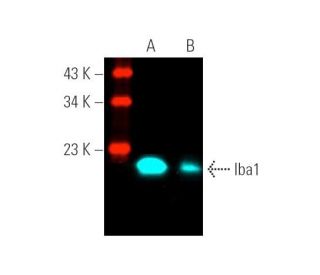
Date published: 2025-9-7

021-6093-6350

SCBT Portrait Logo
Seach Input

Iba1 抗体 (1022-5): sc-32725

3.9(38)
寫評論提問

说明书
  • Iba1 抗体 1022-5 是小鼠单克隆 IgG2b κ,Iba1抗体, 在219篇文献中引用,规格为200 µg/ml
  • 重组human同种异体炎症因子-1
  • Iba1 抗体 (1022-5) 推荐用于 WB, IP, IF, IHC(P) 和 FCM,检测mouse, rat 和human 来源的 Iba1
  • 抗Iba1抗体(1022-5)可与琼脂糖结合用于IP;与HRP结合用于WB、IHC(P)和ELISA;与藻红蛋白FITC结合用于IF、IHC(P)和FCM
  • 还可偶联Alexa Fluor® 488, Alexa Fluor® 546, Alexa Fluor® 594Alexa Fluor® 647,用于WB (RGB), IF, IHC(P) 和 FCM, 以及用于RGB荧光成像系统,例如iBright™ FL1000, FluorChem™, Typhoon, Azure和其他类似的系统
  • 还可偶联Alexa Fluor® 680Alexa Fluor® 790, 用于WB (NIR), IF 和 FCM; 以及用于近红外(NIR)检测系统,如LI-COR®/Odyssey®, iBright™ FL1000, FluorChem™, Typhoon, Azure和类似系统
  • 关于如何获取Iba1 (1022-5): sc-32725免费10 µg小样,联系我们技术服务部门 (或者您当地的代理商)了解详情。
  • m-IgG Fc BP-HRP是Iba1 Antibody (1022-5) 适用于 WB 和 IHC(P) 应用。 的首选辅助检测试剂。该试剂现与Iba1 Antibody (1022-5) 搭配使用(请参阅下面的订购信息)。

快捷链接

関連項目

Iba1抗体(1022-5)是一种小鼠单克隆IgG2b kappa轻链抗体,可通过蛋白质印迹(WB)、免疫沉淀(IP)、免疫荧光(IF)、免疫组织化学和流式细胞术(FCM)检测小鼠、大鼠和人源Iba1蛋白。Iba1 (1022-5) 抗体有非结合型和多种结合型可供选择,包括琼脂糖、辣根过氧化物酶 (HRP)、藻红蛋白 (PE)、异硫氰酸荧光素 (FITC) 和多种 Alexa Fluor® 结合物。Iba1蛋白,也称为离子钙结合适配体分子1或同种异体炎症因子-1,是一种由147个氨基酸组成的细胞质蛋白,在巨噬细胞活化和功能中起着至关重要的作用,因此Iba1在免疫反应和神经炎症中具有重要意义。从结构上看,Iba1包含两个EF-hand结构域,可促进钙结合,这对于细胞信号传导通路的功能至关重要。在细胞因子和干扰素的刺激下,Iba1从与肌动蛋白的初始共定位转移到板状足突,这突出了其在细胞对损伤的反应中的动态作用。此外,Iba1可作为人类小胶质细胞的标记物,并在受伤的骨骼肌中由巨噬细胞表达,这表明其在中枢神经系统和外周组织中的重要性。编码Iba1的基因位于6p21.33染色体上肿瘤坏死因子基因簇内,该基因簇属于人类主要组织相容性复合体区域的一部分,进一步强调了其在免疫调节和疾病进程中的重要性。

仅限研究使用。不适用于诊断和治疗用途。

Alexa Fluor® 是Molecular Probes Inc., OR., USA的商标

LI-COR®和 Odyssey® 是LI-COR Biosciences的注册商标

Iba1 抗体 (1022-5) 参考文献:

  1. 人类 MHC 中靠近 TNF 基因座的新基因的预测产物中包括一个 Ig 超家族新成员和一个 V-ATPase G 亚基。  |  Neville, MJ. and Campbell, RD. 1999. J Immunol. 162: 4745-54. PMID: 10202016
  2. AIF-1 是一种肌动蛋白聚合反应和 Rac1 激活蛋白,可促进血管平滑肌细胞迁移。  |  Autieri, MV., et al. 2003. Circ Res. 92: 1107-14. PMID: 12714565
  3. 小胶质细胞/巨噬细胞特异性蛋白 Iba1 与 fimbrin 结合并增强其肌动蛋白束缚活性。  |  Ohsawa, K., et al. 2004. J Neurochem. 88: 844-56. PMID: 14756805
  4. 高密度 SNP 基因分型确定了高加索人群 TNF Block 的 17 个不同单倍型:对单倍型标记的影响。  |  Allcock, RJ., et al. 2004. Hum Mutat. 24: 517-25. PMID: 15523649
  5. AIF-1 转基因小鼠平滑肌细胞对损伤的活化和新生内膜形成增加。  |  Sommerville, LJ., et al. 2008. Arterioscler Thromb Vasc Biol. 28: 47-53. PMID: 17991871
  6. 同种异体炎症因子-1能增强巨噬细胞的吞噬活性,并加速载脂蛋白E-/-小鼠动脉粥样硬化的进展。  |  Mishima, T., et al. 2008. Int J Mol Med. 21: 181-7. PMID: 18204784
  7. AIF-1 的表达调控着内皮细胞的活化, 信号转导和血管生成。  |  Tian, Y., et al. 2009. Am J Physiol Cell Physiol. 296: C256-66. PMID: 18787073
  8. 人类小胶质细胞中HLA-DR、Iba1和CD68的染色揭示了部分重叠的表达,这取决于细胞形态和病理。  |  Hendrickx, DAE., et al. 2017. J Neuroimmunol. 309: 12-22. PMID: 28601280
  9. 在小鼠大脑中,使用IBA1和TMEM119进行免疫荧光染色,用于分析小胶质细胞密度、形态和外周髓样细胞浸润。  |  González Ibanez, F., et al. 2019. J Vis Exp.. PMID: 31710033
  10. 阿尔茨海默病中小胶质细胞标记物Iba1、TMEM119和P2RY12的共表达模式。  |  Kenkhuis, B., et al. 2022. Neurobiol Dis. 167: 105684. PMID: 35247551
  11. 人异体炎症因子-1 的 cDNA 克隆:受伤大鼠颈动脉的组织分布, 细胞因子诱导和 mRNA 表达。  |  Autieri, MV. 1996. Biochem Biophys Res Commun. 228: 29-37. PMID: 8912632
  12. IRT-1,一种编码抑制生长的碱性亮氨酸拉链蛋白的新型伽马干扰素响应转录本。  |  Autieri, MV. and Agrawal, N. 1998. J Biol Chem. 273: 14731-7. PMID: 9614071

订购信息

产品名称产品编号规格价格数量收藏夹

Iba1 抗体 (1022-5)

sc-32725
200 µg/ml
RMB2377.00

Iba1 (1022-5): m-IgG Fc BP-HRP 套装

sc-525535
200 µg Ab; 10 µg BP
RMB2662.00

Iba1 抗体 (1022-5) AC

sc-32725 AC
500 µg/ml, 25% agarose
RMB3129.00

Iba1 抗体 (1022-5) HRP

sc-32725 HRP
200 µg/ml
RMB2377.00

Iba1 抗体 (1022-5) FITC

sc-32725 FITC
200 µg/ml
RMB2482.00

Iba1 抗体 (1022-5) PE

sc-32725 PE
200 µg/ml
RMB2580.00

Iba1 抗体 (1022-5) Alexa Fluor® 488

sc-32725 AF488
200 µg/ml
RMB2685.00

Iba1 抗体 (1022-5) Alexa Fluor® 546

sc-32725 AF546
200 µg/ml
RMB2685.00

Iba1 抗体 (1022-5) Alexa Fluor® 594

sc-32725 AF594
200 µg/ml
RMB2685.00

Iba1 抗体 (1022-5) Alexa Fluor® 647

sc-32725 AF647
200 µg/ml
RMB2685.00

Iba1 抗体 (1022-5) Alexa Fluor® 680

sc-32725 AF680
200 µg/ml
RMB2685.00

Iba1 抗体 (1022-5) Alexa Fluor® 790

sc-32725 AF790
200 µg/ml
RMB2685.00

请问生产日期是多久啊

Asked by: wwxl
感谢您的提问。您可以拨打电话:021-60936350,发送邮件到[email protected],或者我们官网上的在线聊天,联系我们的亚洲技术支持团队,了解更多信息。 Thank you for your question. Please contact our Technical Service Department for more information. Our Asian Technical Service team is available at (+86) 021-60936350 or [email protected] or by live chat on our website.
Answered by: Jiajun Z
Date published: 2023-07-27

Hello, I am interested to see human macrophages in tumour xenogratfs from NSG mice. Do you suggest anti-Iba1 antibody to suit my objective? What if I have to see mouse macrophages as well ? Is there any other marker to differentiate both ?

Asked by: SANJEEV
Thank you for your question. It would be helpful if you could call us directly at 800-457-3801 ext.1, allowing for a more interactive discussion of this and other related questions.
Answered by: Blake J
Date published: 2021-08-16

请问,我使用此抗体对小鼠脊髓进行免疫组化染色,神经元为什么会呈现强阳性?

Asked by: PURPLE
感谢您的提问。请联系我们的技术支持部门。亚洲技术支持电话(+86) 021-60936350,邮箱[email protected],或选择网页上Live Chat中的“中文支持”。 Thank you for your inquiry. Please contact our Technical Service Department. Our Asian Technical Service team is available at (+86) 021-60936350 or [email protected] or by live chat on our website.
Answered by: Technical Support 9
Date published: 2022-03-27

I use the IBA-1 antibody (1022-5): sc-32725 for brain tissue of rats,but my band about 200kd,while it at 20kd band, so I don't how to explain.

Asked by: yuyu
Thank you for your question. The expected molecular weight of Iba1 is approximately 17 kDa. Please get in touch with our technical service department for troubleshooting advice on your experiment.
Answered by: Tech Support Europe
Date published: 2020-12-31

hi,i bought the IBA-1 antibody several days ago.the product number is SC32725. i use it for IF.But it seem that the antibody do not work .i need technical support,thank you

Asked by: Anonymous
Thank you for your question. We are sorry to hear that there is an issue with this antibody. Please contact [email protected] so we can assist you in troubleshooting your experiment.
Answered by: Tech Support Europe
Date published: 2020-08-04

Can I use the IBA 1 antibody (which one) for staining of paraffin embedded pig tissue?

Asked by: Anni1703
Thank you for your question. This antibody has not been tested for porcine use, and is not recommended or warrantied for this use. It is recommended for IHC using paraffin embedded tissue sections. If you have any further questions, please contact Technical Service.
Answered by: Tech Service
Date published: 2019-04-30

Dear I would to receive the data sheet the item 32725 IBA1 antibody Alexa 488 Sincerally Dr Gustavo

Asked by: Argañaraz
Thank you for your question. We do not provide separate product data sheets for the conjugated versions of this antibody. Please contact our Technical Service Department for further support.
Answered by: Technical Support
Date published: 2019-02-23

Can I use this mouse monoclonal antibody for immunohistochemical staining of mouse tissue?

Asked by: DefinitelyNotMatt
Thank you for your question. Yes. We recommend using a directly conjugated mouse monoclonal primary antibody. This mouse monoclonal antibody is currently available conjugated to phycoerythrin (sc-32725 PE) or fluoroscein (sc-32725 FITC), Alexa Fluor® 488 (sc-32725 AF488) or Alexa Fluor® 647 (sc-32725 AF647). Please contact Technical Service if you have further questions concerning this product.
Answered by: Tech Service 9
Date published: 2016-12-30
  • y_2025, m_9, d_7, h_4CST
  • bvseo_bulk, prod_bvqa, vn_bulk_3.0.42
  • cp_1, bvpage1
  • co_hasquestionsanswers, tq_8
  • loc_zh_CN, sid_32725, prod, sort_[SortEntry(order=LAST_APPROVED_ANSWER_SUBMISSION_TIME, direction=DESCENDING)]
  • clientName_scbt
  • bvseo_sdk, java_sdk, bvseo-4.0.0
  • CLOUD, getContent, 112ms
  • QUESTIONS, PRODUCT
Rated 5 out of 5 by from it works well in immunofluorescenceWe used it in dection of microglial activation in the retina tissues. The results of immunofluorescence shows intensive signal and little background signals.
Date published: 2024-01-16
Rated 5 out of 5 by from ottimo per wbtestato su macrofagi umani in wb. Molto soddisfatta
Date published: 2023-03-10
Rated 5 out of 5 by from SK030204V100This antibody is well-cited for a variety of applications including WB, IP, IF, and even ELISA. Strong, clear signal with little to no background. Works well in a variety of species including mouse, rat, and human samples.
Date published: 2022-01-05
Rated 5 out of 5 by from 东西不错This antibody is well-cited for a variety of applications including WB, IP, IF, and even ELISA. Strong, clear signal with little to no background. Works well in a variety of species including mouse, rat, and human samples.
Date published: 2022-01-05
Rated 5 out of 5 by from 染色(IHC)マウス脳に対して 1:100、TMEM119抗体との共染色実施。 二次抗体はDyLight 488使用。 良好な染色が得られた。
Date published: 2021-12-17
Rated 4 out of 5 by from Iba signal in lung macrophagesIba1 was detected on mice tumor lung paraffin sections via the techonology of IF. Dilution 1: 100. I think this antibody could dilute. We use heat and citrate buffer for antigen retrieval. WAKO Iba1 offer better signal for macrophages
Date published: 2020-10-19
Rated 3 out of 5 by from IF on mice brainWe tested this antibody for immunofluorescence to detect IBA-1on microglial cells on mice brain (fixed in PFA). It works well at 1:200 48hours of incubation in PB+0.3% Triton X-100.
Date published: 2020-01-28
Rated 2 out of 5 by from Antibody reviewDid not work in IF staining with mouse tissue. May work with WB.
Date published: 2018-05-29
  • y_2025, m_9, d_7, h_4
  • bvseo_bulk, prod_bvrr, vn_bulk_3.0.42
  • cp_1, bvpage1
  • co_hasreviews, tv_0, tr_38
  • loc_zh_CN, sid_32725, prod, sort_[SortEntry(order=SUBMISSION_TIME, direction=DESCENDING)]
  • clientName_scbt
  • bvseo_sdk, java_sdk, bvseo-4.0.0
  • CLOUD, getReviews, 16ms
  • REVIEWS, PRODUCT
Iba1 抗体 (1022-5) is rated 3.9 out of 5 by 38.
  • y_2025, m_9, d_7, h_4
  • bvseo_bulk, prod_bvrr, vn_bulk_3.0.42
  • cp_1, bvpage1
  • co_hasreviews, tv_0, tr_38
  • loc_zh_CN, sid_32725, prod, sort_[SortEntry(order=SUBMISSION_TIME, direction=DESCENDING)]
  • clientName_scbt
  • bvseo_sdk, java_sdk, bvseo-4.0.0
  • CLOUD, getAggregateRating, 115ms
  • REVIEWS, PRODUCT