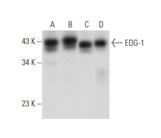
Date published: 2025-9-6

021-6093-6350

SCBT Portrait Logo
Seach Input

EDG-1/S1P1/S1PR1 抗体 (A-6): sc-48356

4.0(13)
寫評論提問

说明书
  • EDG-1/S1P1/S1PR1 抗体 A-6 是小鼠单克隆 IgG3 κ,EDG-1/S1P1/S1PR1抗体, 在23篇文献中引用,规格为200 µg/ml
  • 免疫human物种的EDG-1的氨基酸322-381
  • 推荐用于 human 来源的EDG-1 WB, IP, IF, IHC(P) 和 ELISA检测
  • 另提供琼脂糖偶联抗体,规格为琼脂糖装,可用于IP实验;HRP 偶联抗体用于WB, IHC(P)和ELISA实验;以及藻红蛋白、FITC、Alexa Fluor® 488Alexa Fluor® 647 偶联抗体,规格为用于IF、IHC(P)和FCM实验
  • 关于如何获取EDG-1/S1P1/S1PR1 (A-6): sc-48356免费10 µg小样,联系我们技术服务部门 (或者您当地的代理商)了解详情。
  • 目前,我们还没有完成EDG-1/S1P1/S1PR1 抗体 (A-6)的首选二抗检测试剂的鉴定。这项工作正在进行中。

快捷链接

関連項目

EDG-1 抗体 (A-6) 是一种小鼠单克隆 IgG3 kappa 轻链抗体,可通过免疫印迹 (WB)、免疫沉淀 (IP)、免疫荧光 (IF)、免疫组织化学和酶联免疫吸附试验 (ELISA) 检测人源 EDG-1。EDG-1 (A-6) 抗体有非共轭型和多种共轭型,包括琼脂糖、辣根过氧化物酶 (HRP)、藻红蛋白 (PE)、异硫氰酸荧光素 (FITC) 和多种 Alexa Fluor® 共轭物。EDG-1 又称 S1PR1 或 S1P1,是 G 蛋白偶联受体内皮分化基因(EDG)家族的成员,在介导鞘磷脂信号分子(如鞘氨醇-1-磷酸(S1P)和溶血磷脂酸(LPA))的作用方面起着至关重要的作用。EDG-1 主要位于细胞表面,通过与 Gi 蛋白耦合激活 Akt 等下游信号通路,参与各种细胞过程,包括细胞存活、生长、迁移和分化。这在胶质瘤细胞中尤为重要,因为 EDG-1 的表达与肿瘤的进展和存活有关。EDG-1 与多种 G 蛋白相互作用的能力影响着多种生理反应,使 EDG-1 成为癌症生物学研究和治疗开发的重要靶点。

仅限研究使用。不适用于诊断和治疗用途。

Alexa Fluor® 是Molecular Probes Inc., OR., USA的商标

LI-COR®和 Odyssey® 是LI-COR Biosciences的注册商标

EDG-1/S1P1/S1PR1 抗体 (A-6) 参考文献:

  1. 鞘氨醇1-磷酸受体EDG-1、EDG-3和EDG-5的差异药理学特性和信号转导。  |  Ancellin, N. and Hla, T. 1999. J Biol Chem. 274: 18997-9002. PMID: 10383399
  2. 溶血磷脂和溶血磷脂的 G 蛋白偶联细胞受体亚家族。  |  Goetzl, EJ. and An, S. 1999. Adv Exp Med Biol. 469: 259-64. PMID: 10667339
  3. 1-磷酸肾上腺素是 G 蛋白偶联受体 EDG-6 的配体。  |  Van Brocklyn, JR., et al. 2000. Blood. 95: 2624-9. PMID: 10753843
  4. 通过G蛋白偶联受体EDG-1家族进行鞘氨醇-1-磷酸信号传导。  |  Hla, T., et al. 2000. Ann N Y Acad Sci. 905: 16-24. PMID: 10818438
  5. 鞘氨醇-1-磷酸:G蛋白偶联受体EDG-1家族的配体。  |  Spiegel, S. 2000. Ann N Y Acad Sci. 905: 54-60. PMID: 10818441
  6. 哺乳动物细胞中的 1-磷酸肾上腺素信号传导  |  Pyne, S. and Pyne, NJ. 2000. Biochem J. 349: 385-402. PMID: 10880336
  7. Edg-1和Edg-5(1-磷酸鞘氨醇受体)在C6胶质瘤细胞信号通路中的不同作用。  |  Sato, K., et al. 2000. Brain Res Mol Brain Res. 85: 151-60. PMID: 11146117
  8. 溶血磷脂酸受体对 Jurkat T 细胞通过 Matrigel 模型基底膜迁移的选择性影响  |  Zheng, Y., et al. 2001. J Immunol. 166: 2317-22. PMID: 11160288
  9. Edg2 受体在成年大鼠大脑中的分布。  |  Handford, EJ., et al. 2001. Neuroreport. 12: 757-60. PMID: 11277579
  10. 1- 磷酸鞘氨醇通过内皮细胞中的 Gi 蛋白/磷酸肌醇 3- 激酶途径激活 Akt, 一氧化氮的产生和趋化作用。  |  Morales-Ruiz, M., et al. 2001. J Biol Chem. 276: 19672-7. PMID: 11278592
  11. N-糖基化在鞘氨醇1-磷酸刺激细胞中Edg-1/S1P1动态变化中的作用。  |  Kohno, T. and Igarashi, Y. 2004. Glycoconj J. 21: 497-501. PMID: 15750791

订购信息

产品名称产品编号规格价格数量收藏夹

EDG-1/S1P1/S1PR1 抗体 (A-6)

sc-48356
200 µg/ml
RMB2377.00

EDG-1/S1P1/S1PR1 抗体 (A-6) AC

sc-48356 AC
500 µg/ml, 25% agarose
RMB3129.00

EDG-1/S1P1/S1PR1 抗体 (A-6) Alexa Fluor® 488

sc-48356 AF488
200 µg/ml
RMB2685.00

EDG-1/S1P1/S1PR1 抗体 (A-6) Alexa Fluor® 647

sc-48356 AF647
200 µg/ml
RMB2685.00

EDG-1/S1P1/S1PR1 抗体 (A-6) FITC

sc-48356 FITC
200 µg/ml
RMB2482.00

EDG-1/S1P1/S1PR1 抗体 (A-6) HRP

sc-48356 HRP
200 µg/ml
RMB2377.00

EDG-1/S1P1/S1PR1 抗体 (A-6) PE

sc-48356 PE
200 µg/ml
RMB2580.00

If I Stored the antibody at -20° C, does it affect its activity?

Asked by: June 0615
Thank you for your question. EDG-1/S1P1/S1PR1 抗体 (A-6): sc-48356 should be stored at 4° C, do not freeze. If it is stored at -20° C, it may affect its activity.
Answered by: Technical Service SH
Date published: 2022-02-28

What is the recommended secondary detection reagent to be used with the EDG-1 Antibody (A-6): sc-48356 for Western blot?

Asked by: Trav11
Thank you for your question. We recommend using one of our exclusive Mouse IgG Binding Proteins as a secondary detection reagent, such as m-IgGκ BP-HRP: sc-516102, for Western blot. A complete list of all available binding proteins is available on our website here: https://www.scbt.com/scbt/browse/support-products-mouse-igg-binding-proteins/_/N-ecrety If you have any further questions or concerns, please feel free to contact our Technical Service department by calling 800-457-8301 option 2, emailing [email protected], or using the Live Chat function on our website.
Answered by: Technical Support 15
Date published: 2017-01-22
  • y_2025, m_9, d_6, h_5CST
  • bvseo_bulk, prod_bvqa, vn_bulk_3.0.42
  • cp_1, bvpage1
  • co_hasquestionsanswers, tq_2
  • loc_zh_CN, sid_48356, prod, sort_[SortEntry(order=LAST_APPROVED_ANSWER_SUBMISSION_TIME, direction=DESCENDING)]
  • clientName_scbt
  • bvseo_sdk, java_sdk, bvseo-4.0.0
  • CLOUD, getContent, 109ms
  • QUESTIONS, PRODUCT
Rated 4 out of 5 by from Works wellDoesn't seem to give a super robust signal, but it does work. We like to try and re-use antibodies for Westerns, however this antibody seems to only work when fresh, at a 1:250 dilution.
Date published: 2019-01-14
Rated 5 out of 5 by from worked well1:50 dilution for IF with human brain section, worked well ,expected staining.
Date published: 2018-05-26
Rated 5 out of 5 by from Working very good for ImmunoblottingI bought a free sample of this antibody. It is working excellent. Clean band.
Date published: 2017-09-18
Rated 5 out of 5 by from Detects GFP tagged and endogenous EDG-1 in IFAntibody detects successfully GFP tagged and endogenous EDG-1 in human cell lines.
Date published: 2017-08-30
Rated 1 out of 5 by from Did not work in mouse tissueI received a free sample of this because H-60 has been discontinued. I tested in both IHC and Western. There was no staining in IHC and the Western showed no bands. I have attached a blot from the H-60 antibody that I have that is >4 years old. I was really hoping the monoclonal would work. I used the same tissue and protocol for both Westerns. Santa Cruz person I worked with was great, no complaints there.
Date published: 2017-07-05
Rated 1 out of 5 by from Not good for immunohistochemistryWe obtained a sample of this antibody to test since the EDG-1 (H-60) is no longer available. The signal was not above the background signal and it did not label any structures in the cell specifically when compared to our old antibody.
Date published: 2017-06-15
Rated 5 out of 5 by from Great productVery nice western blot data, strong, clear signal; also strong IHC staining results
Date published: 2017-02-02
Rated 5 out of 5 by from Excellent!This antibody works great in western blot analysis of EDG-1 expression in human brain tissue extract.
Date published: 2017-01-11
  • y_2025, m_9, d_6, h_5
  • bvseo_bulk, prod_bvrr, vn_bulk_3.0.42
  • cp_1, bvpage1
  • co_hasreviews, tv_0, tr_13
  • loc_zh_CN, sid_48356, prod, sort_[SortEntry(order=SUBMISSION_TIME, direction=DESCENDING)]
  • clientName_scbt
  • bvseo_sdk, java_sdk, bvseo-4.0.0
  • CLOUD, getReviews, 30ms
  • REVIEWS, PRODUCT
EDG-1/S1P1/S1PR1 抗体 (A-6) is rated 4.0 out of 5 by 13.
  • y_2025, m_9, d_6, h_5
  • bvseo_bulk, prod_bvrr, vn_bulk_3.0.42
  • cp_1, bvpage1
  • co_hasreviews, tv_0, tr_13
  • loc_zh_CN, sid_48356, prod, sort_[SortEntry(order=SUBMISSION_TIME, direction=DESCENDING)]
  • clientName_scbt
  • bvseo_sdk, java_sdk, bvseo-4.0.0
  • CLOUD, getAggregateRating, 110ms
  • REVIEWS, PRODUCT